Figure 1.
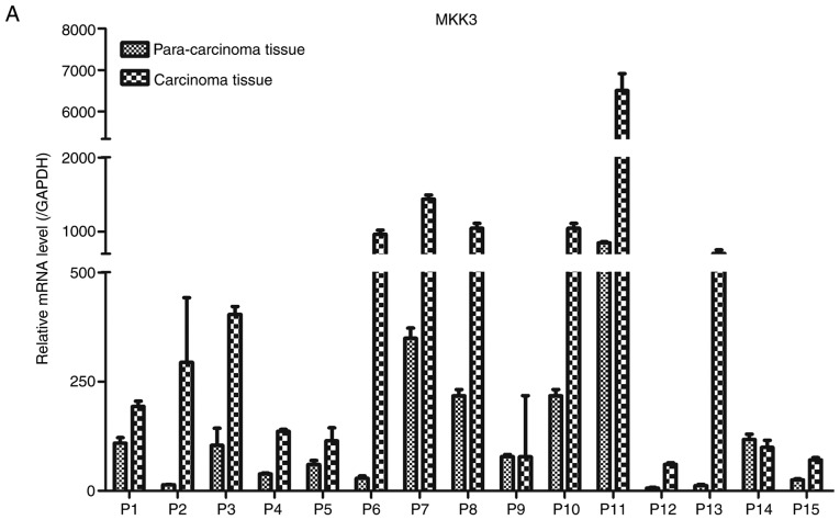
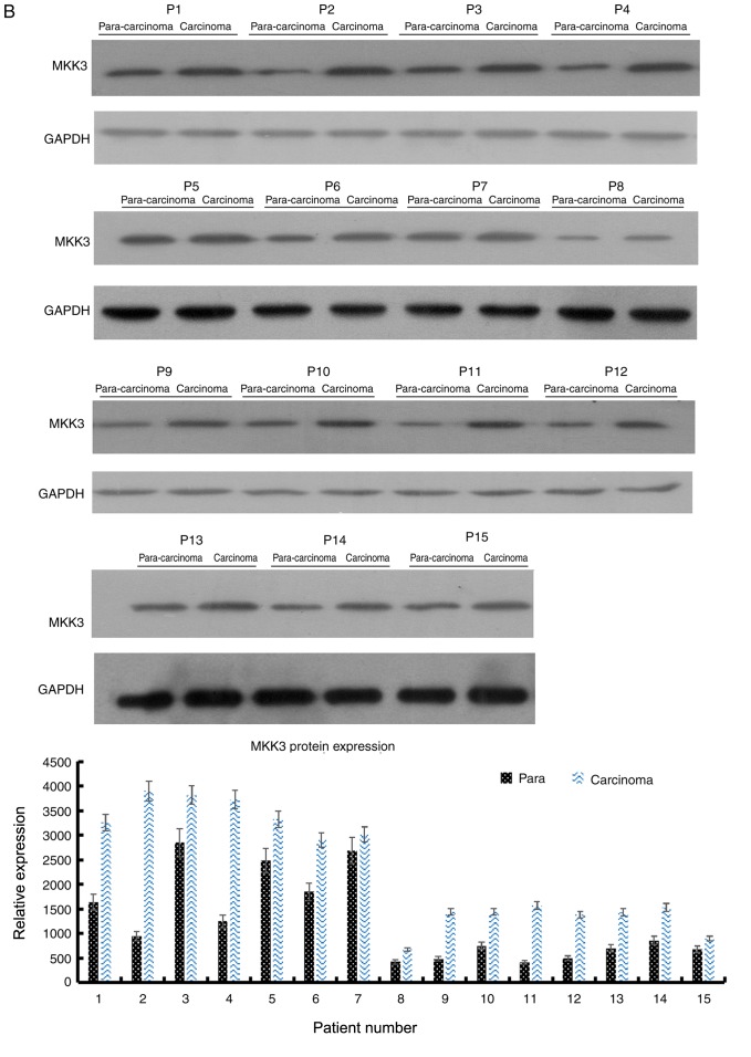
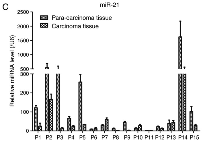
MKK3 and miR-21 expression levels are dysregulated in melanoma tissues. (A) RT-qPCR analysis of MKK3 mRNA levels in 15 pairs of melanoma tissues and adjacent normal tissues. (B) Western blot analysis of MKK3 protein levels in 15 pairs of melanoma tissues and adjacent normal tissues. The band intensity was determined to quantify protein expression levels. MKK3 and miR-21 expression levels are dysregulated in melanoma tissues. (C) RT-qPCR analysis of miR-21 levels in 15 pairs of melanoma tissues and adjacent normal tissues. The results are presented as the mean ± standard deviation of three replicates per tissue sample. MKK3, mitogen-activated protein kinase kinase 3; miR-21, microRNA-21; RT-qPCR, reverse transcription-quantitative PCR; P, patient.


