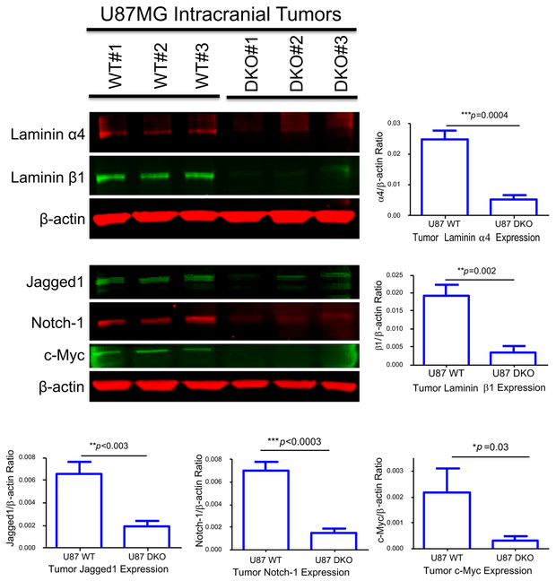
Fig. 4. Characterization of intracranial tumors derived from both WT and laminin-411 DKO U87MG cells by western blot.
Intracranial tumors were dissected from mice inoculated with either WT or the laminin-411 DKO U87MG cells. Tumor samples were collected from three animals per group at the endpoint of their survival and total lysates were used for western blotting analysis. Upper blot: Levels of laminin-411 α4 and β1 in U87MG tumors (WT vs. laminin-411 DKO. Compared to the WT U87MG tumors, the laminin-411 α4 and β1 levels remained significantly lower in DKO tumors; Lower blot: Levels of CSC markers in the tumor samples. Notch-1, Jagged1 as well as the CSC regulator c-Myc are significantly lower than in the WT tumors.

