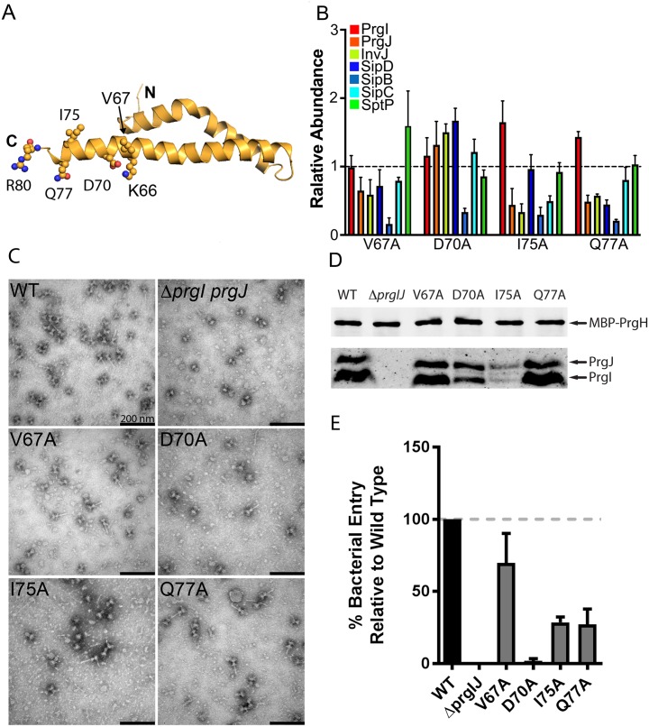
Fig 5. Secretion defects associated with C-terminal residues reveals a complex role for the lumen of the needle filament in type III secretion.
(A) Representation of the location of relevant residues within the atomic structure of PrgI. (B) Bar graph detailing the secretion phenotypes of S. Typhimurium expressing the indicated PrgI mutants. The relative abundance of the secreted substrates has been standardized relative to WT, which was given a value of 1 and is demarcated by a gray dashed line. All values represent the mean ± the standard deviation of three independent experiments. The data was compiled from the data presented in S2 Fig. The underlying data for this panel can be found in S5 Data. (C and D) Electron micrographs (C) and western blot analysis (D) of affinity purified needle complexes obtained from S. Typhimurium strains expressing the indicated PrgI mutants. Samples for the western blot analysis were standardized based on the amount of PrgH. (E) Cultured epithelial cell invasion ability of S. Typhimurium strains expressing the indicated PrgI mutants. Numbers represent the percentage of the inoculum that survived antibiotic treatment due to internalization and are the mean ± standard deviation of three independent experiments normalized to WT, which was set to 100%. The underlying data for this panel can be found in S6 Data. WT, wild-type.

