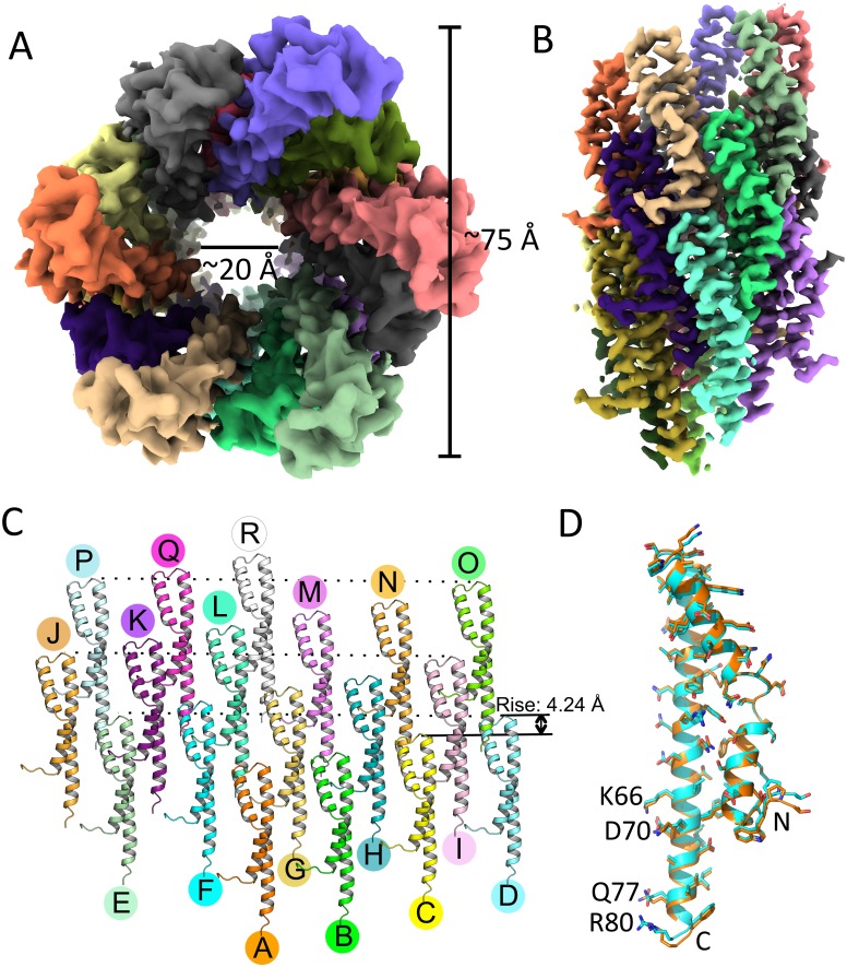
Fig 7. Cryo-EM reconstruction of PrgI needles attached to the base.
(A and B) Top (A) and side (B) views of the PrgI needle. The outer diameter is approximately 75 Å, and the inner diameter is approximately 20 Å. PrgI subunits are rendered in different colors. (C) The PrgI subunits in the needle structure arranged in roughly three turns are shown. (D) Superimposition of a single PrgI subunit extracted from the needle structure attached to the base (cyan), with a PrgI subunit extracted from the structure of isolated PrgI filaments removed from the base (PDB: 6DWB; orange). cryo-EM; PDB, Protein Data Bank.

