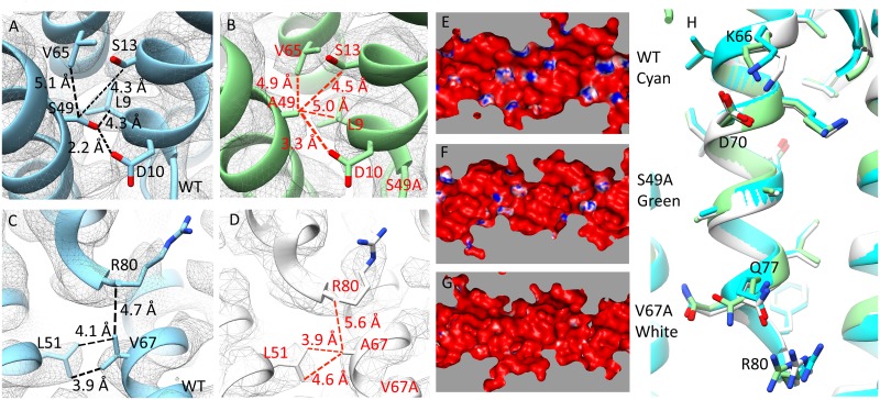
Fig 8. The cryo-EM structures of PrgI mutant proteins revealed structural differences.
(A–D) Hydrogen bonds and hydrophobic interactions of S49 and V67 in WT needle filament and PrgIS49A and PrgIV67A filaments. (E–G) Electrostatic potential maps of the lumen of PrgIWT, PrgIS49A, and PrgIV67A filaments. (H) Cross-section view of the PrgI filaments lumen alignment (PrgIWT [cyan], PrgIS49A [green], and PrgIV67A [white]). cryo-EM, cryo electron microscopy; WT, wild-type.

