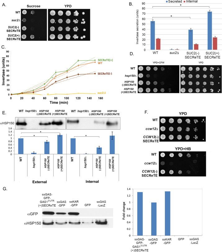
Fig 5. The levels of secretion of endogenous and exogenous proteins are affected by SECReTE strength.
(A) SECReTE enhances the ability to grow on sucrose. The ability of WT, suc2Δ, SUC2(+)SECReTE and SUC2(-)SECReTE yeast to grow on sucrose was examined by drop-test. Cells were grown to mid-log on glucose-containing YPD medium, prior to serial dilution and plating onto sucrose-containing synthetic medium or YPD. Cells were grown for 2 days prior to photo-documentation. The SUC2(-)SECReTE mutant exhibited reduced growth than WT cells, while SUC2(+)SECReTE cells exhibited better growth. suc2Δ cells were unable to grow on sucrose-containing medium. (B) SECReTE enhances invertase secretion. The indicated strains from A were analyzed using an invertase secretion assay. Both internal and secreted invertase activity was measured in units (1 U = 1 μmol glucose released/min per O.D.600 unit) after glucose de-repression. Both activities were reduced in SUC2(-)SECReTE cells and elevated in SUC2(+)SECReTE cells. Error bars represent the standard deviation from three experimental repeats. *p<0.0161 (t-test). (C) SECReTE presence enhances the rate of invertase secretion. The indicated strains from A were incubated in low glucose medium for varying times (0-150min) and analyzed using the invertase secretion assay, as in B. Secreted invertase activity was measured in units (1 U = 1 μmol glucose released/min per O.D.600 unit) after glucose de-repression. Error bars represent the standard deviation from three experimental repeats. Linear regression of the data (dotted lines) from inverse reciprocal plots was used to determine the T1/2 for half-maximal accumulation and rate; “x” mark on line indicates time of half-maximal accumulation. The rate of invertase secretion is enhanced in (+)SECReTE cells and reduced in (-)SECReTE cells relative to WT. (D) SECReTE enhances the ability to grow on calcofluor white. The ability of WT, hsp150Δ, HSP150(+)SECReTE and HSP150(-)SECReTE cells to grow on CFW was examined by drop-test. Cells were grown to mid-log on YPD, prior to serial dilution and plating on YPD alone or YPD plates containing CFW, and incubated at 30°C. Cells were grown for 2 days prior to photodocumentation. The HSP150(-)SECReTE mutant exhibited hypersensitivity in comparison to WT cells, while HSP150(+)SECReTE cells were less sensitive. hsp150Δ cells grew poorly on medium containing CFW. (E) SECReTE enhances Hsp150 secretion. The indicated strains from D were subjected to the Hsp150 secretion assay. Cells were grown to mid-log phase at 37°C for 4hrs and examination in cell lysates (internal) or medium (external) by Western analysis using anti-Hsp150 antibodies. External Hsp150 was decreased in HSP150(-)SECReTE cells in comparison to WT, while it was increased in the HSP150(+)SECReTE strain. Internal Hsp150 was decreased in HSP150(-)SECReTE cells and also slightly in HSP150(+)SECReTE cells, in comparison with WT cells. No internal nor external Hsp150 was detected in the lysate or medium derived from hsp150Δ cells, respectively. Band intensity was quantified using ImageJ and presented in the histogram below. A representative experiment is shown in the top panels for both external and internal Hsp150 secretion. The graphs below represent the ratio of the intensity of all samples relative to that of WT for three biological repeats, *p<0.05 (t-test). Error bars represent the standard deviation. (F) SECReTE enhances the ability to grow on hygromycin B. The ability of WT, ccw12Δ, and CCW12(-)SECReTE cells to grow on HB was examined by drop-test. Cells were grown to mid-log on glucose-containing YPD medium, prior to serial dilution and plating onto HB-containing YPD or YPD alone. Cells were grown for 2 days prior to photodocumentation. The CCW12(-)SECReTE strain was more sensitive to HB stress in comparison to WT cells. ccw12Δ cells were unable to grow on medium containing HB. (G) SECReTE enhances secretion of an exogenous protein, SSGAS1-GFP. Yeast expressing SSGAS1-GFP-3’GAS1UTR(+)SECReTE, SSGAS1-GFP, SSKAR2-GFP, GFP, and SSGAS1-LacZ from plasmids were grown to mid-log phase on synthetic medium containing 2% raffinose and shifted to 3% galactose-containing medium for 4hrs. Proteins expressed from the different strains were TCA precipitated from the medium and the precipitates resolved by SDS-PAGE. GFP was detected with an anti-GFP antibody, while Hsp150 was detected with an anti-Hsp150 antibody and was used as a loading control. Band intensity was quantified using ImageJ; intensity was scored relative to SSGAS1-GFP secretion. Addition of the GAS1 3’UTR mutated to contain SECReTE improved the secretion of SS-Gas1 and was comparable to that of SSKAR2-GFP. GFP lacking a signal sequence (GFP) was not secreted and SSGAS1-LacZ was used as a negative control for GFP detection.

