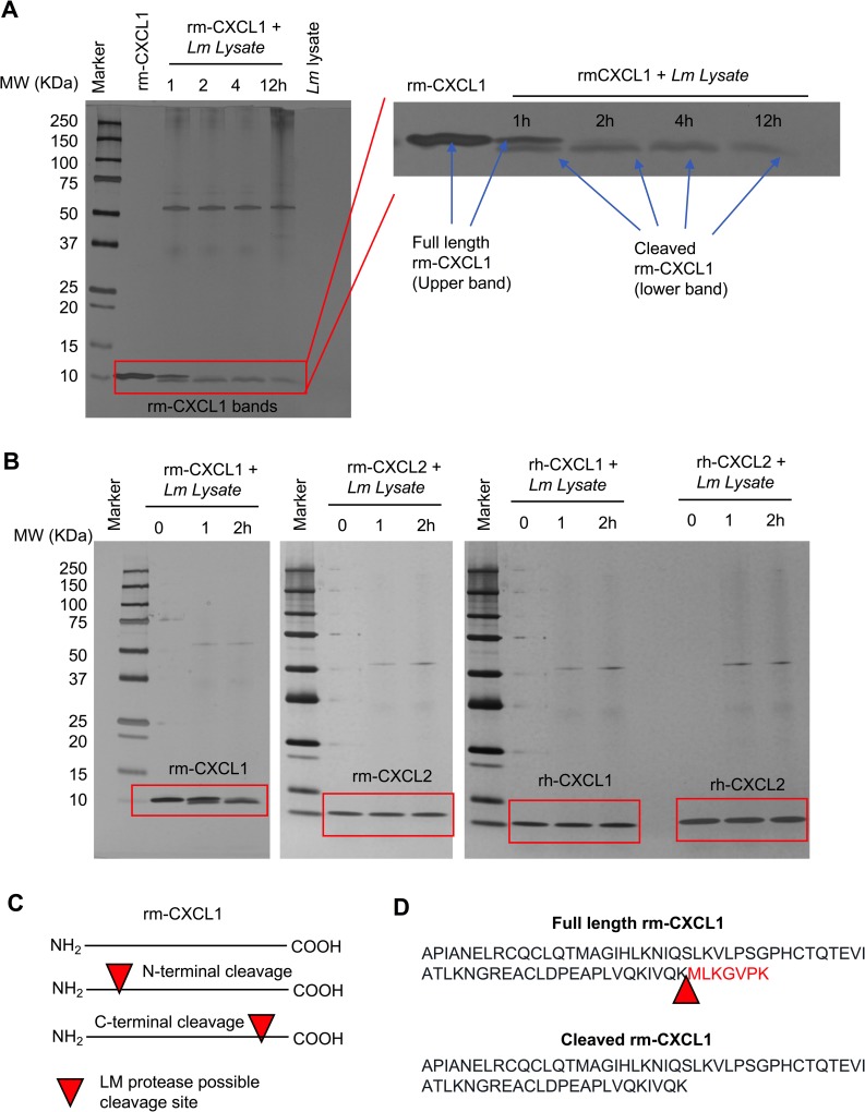
Fig 5. L. major cleaves CXCL1 at the C-terminal end.
(A) A silver-stained SDS-PAGE demonstrate time-dependent cleavage of rm-CXCL1. Rm-CXCL1 were left alone or treated with Lm lysate for the indicated time points. Rm-CXCL1 bands were enlarged to show the cleavage products on right. (B) Indicated chemokines were treated with Lm lysate for indicated time-points and subjected to SDS-PAGE followed by silver-staining. (C) Hypothesized L. major cleavage site on rm-CXCL1 based on the observed cleaved rm-CXCL1 bands in (A and B). Arrow denotes possible cleavage site on CXCL1 (D) Identification of CXCL1 cleavage site by Mass Spectrometry analysis (raw data presented in S4 Fig). Arrow represents cleavage site on rm-CXCL1 targeted by L. major. Data in A and B are representative of at least three independent experiments.

