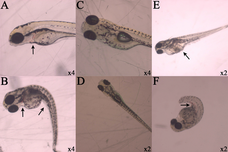
Figure 5.
Observed lesions in zebrafish larvae. Respective magnifications are displayed in the lower right hand corner of each photograph, and black arrows indicate lesions of interest. Cisplatin exposure at 30 mg/L demonstrate pericardial sac hemorrhaging (A and B) and spinal curvature (B) after dechorionation. Control organisms without lesions are centered (C and D). PMC79 exposure at arrows indicate yolk sac edema and protein coagulation and precipitation (E) and hemorrhaging along caudal vein or tail artery (F).

