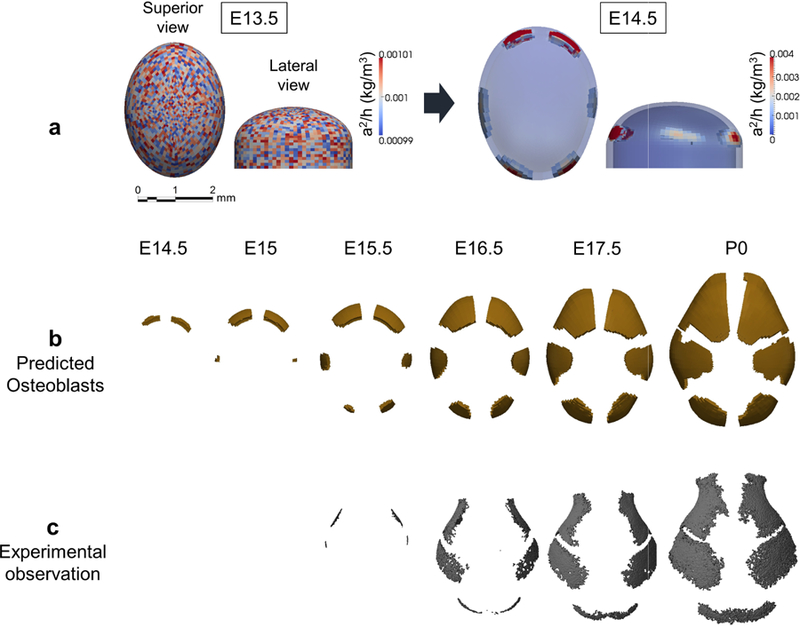
Fig. 3.

The RDE model predicts the location of primary centers of ossification and pattern of cranial vault bone growth. a Computational result of distribution of concentration of activator relative to inhibitor (a2/h) at E13.5 and E14.5. In superior view, anterior at top; in lateral view, anterior is left. b Computational prediction of distribution of differentiating osteoblasts and cranial vault bone formation by embryonic day. Superior view of skulls, anterior at top, posterior at bottom. Ossification centers for right and left frontal bones appear first (~ E14.5), followed by right and left parietal bones (~E15). Two more ossification centers representing the interparietal bone appear at E15.5. c Observed cranial vault bone formation and growth in embryonic mice (see Appendix A).
