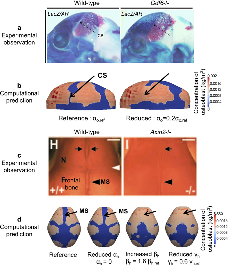
Fig. 6.

Comparison of experimental observations and predictions by the RDE model about cranial dysgenesis due to molecular variants. a Experimental observation of vault bones (stained by alizarin red) of a wild-type (WT) and Gdf 6−/− mouse. Lateral view, rostrum to left, eye at base of coronal suture (CS). CS is open in WT and closed in Gdf 6−/− mouse (adopted from Clendenning and Mortlock (2012)). b Computational prediction of distribution of osteoblasts with the reference value (left, CS patent) and the reduced value of αo (right, CS closed). c Experimental observation of alizarin-red stained cranial vault of WT and Axin2−/− mice (superior view, rostrum at top). Inter-nasal suture between arrows at top; metopic suture (MS) between frontal bones at bottom (adopted from Yu et al. (2005)). d Computational prediction of distribution of osteoblasts, forming bone, and morphology of MS with the reference value (far left), reduced value of αh, increased value of βh, and reduced value of γh.
