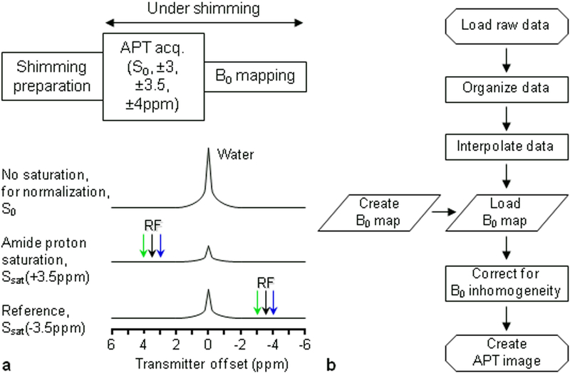
Figure 3.
a: Six-offset APT data acquisition protocol with shimming typically up to the second order. During the APT data acquisition, extra offsets (±3, ±4 ppm) are acquired to correct for the residual B0 inhomogeneity. b: APT data processing flow chart. The procedures include the generation of B0 shift map and correction of APT data using B0 map. Reproduced with permission from Zhou et al., Magn Reson Med 2008;60:842–9.

