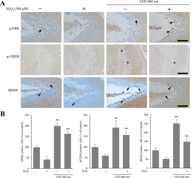
Figure 3.
LED at 660 nm enhances the expression of BDNF in the hippocampus. Hippocampal organotypic slice cultures were subjected to oxidative stress by H2O2 (100 μM), and irradiated with LED at 660 nm. (A) Representative sections of organotypic slices stained with antibodies against p-ERK, p-CREB, and BDNF, as indicated, and counterstained for hematoxylin/eosin. Arrows, asterisks, and arrowheads indicate positively immunostained cells. (B) Quantification of p-ERK-, p-CREB- and BDNF-positive cells in LED-treated hippocampal slices relative to control. The graph shows the percentage of immunostained positive cells when compared to the cells of control hippocampal tissue in which oxidative stress was not induced. NT, not treated; plus and minus symbols indicate the presence or absence of H2O2. **p < 0.01 vs control. Scale bar = 100 µm.

