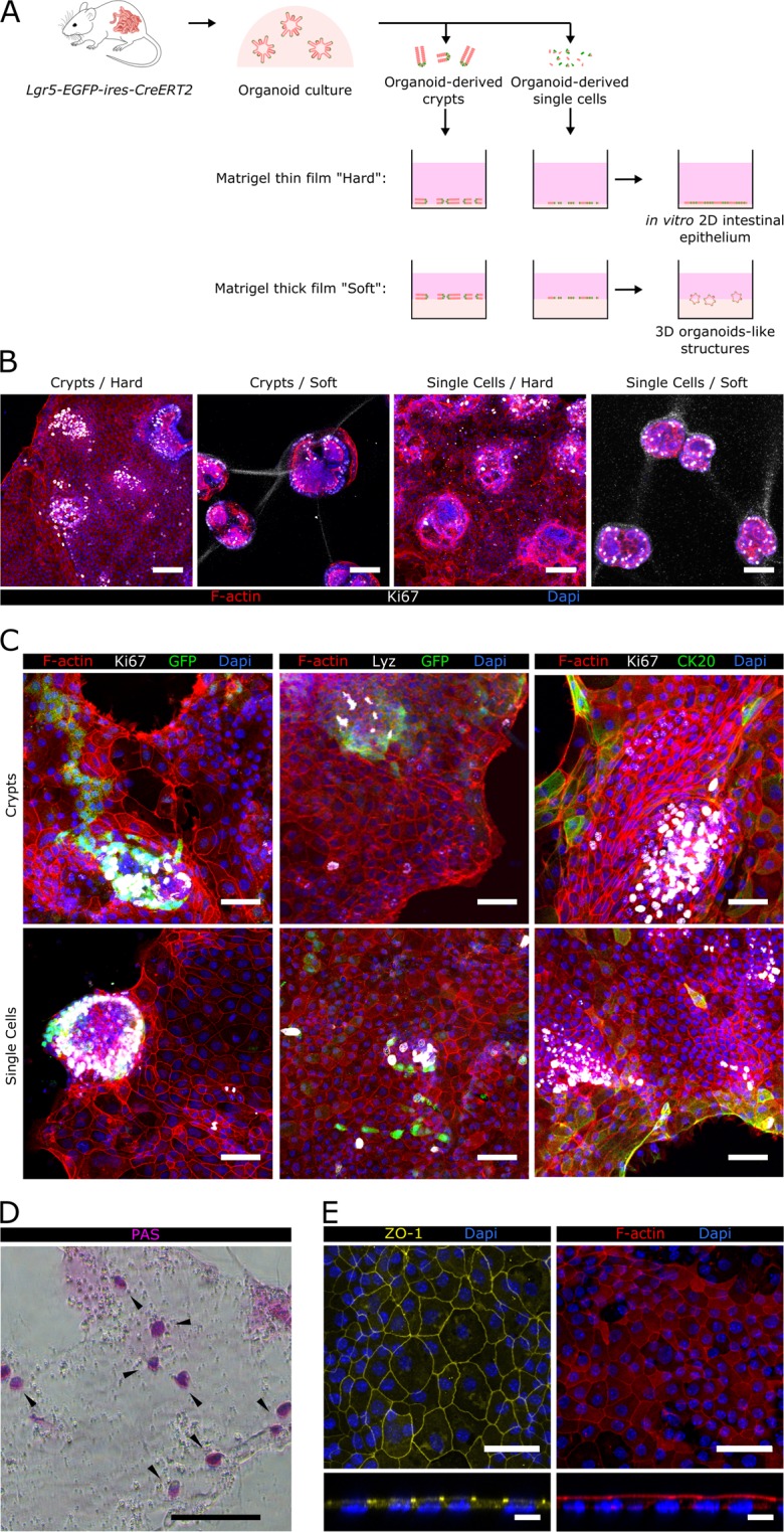
Figure 1.

Epithelial monolayers with in vivo-like cellular organization are formed on Matrigel-coated hard substrates. (A) Scheme depicting the different experimental set-ups employed. Either organoids-derived crypts or single cells where seeded on top of thin or thick layers of Matrigel-coated polystyrene plates. Epithelial structures obtained under each of these conditions are shown. (B) Representative images corresponding to co-immunofluorescence of filamentous actin (F-actin) and Ki67 from organoid-derived crypts and single cells cultured either on a thin film of Matrigel, referred as “Hard” substrate, or on a thick layer of Matrigel, referred as “Soft” substrate after 5 days of culture. Scale bars: 100 µm. (C) Co-immunofluorescence images for GFP (Lgr5-GFP+) and Ki67 (left panel), Lysozyme (Lyz) and GFP (middle panel), and Ki67 and cytokeratin 20 (CK20) (right panel) of epithelial monolayers derived from crypts (upper row) or single cells (lower row) grown on Matrigel-coated hard substrates for 7 days. Scale bars: 50 µm. (D) Image of Periodic acid-Schiff base (PAS) staining of epithelial monolayers grown on Matrigel-coated hard substrates for 7 days. Scale bar: 100 µm. (E) Immunostaining for zonula occludens 1 (ZO-1) and F-actin of epithelial monolayers grown on Matrigel-coated hard substrates for 7 days. Upper and lower panels show the top view and the orthogonal cross-sections of the monolayers, respectively. Scale bars: 50 µm (upper panel); 10 µm (lower panel). In all images Dapi was used to stain the nuclei.
