Figure 5.

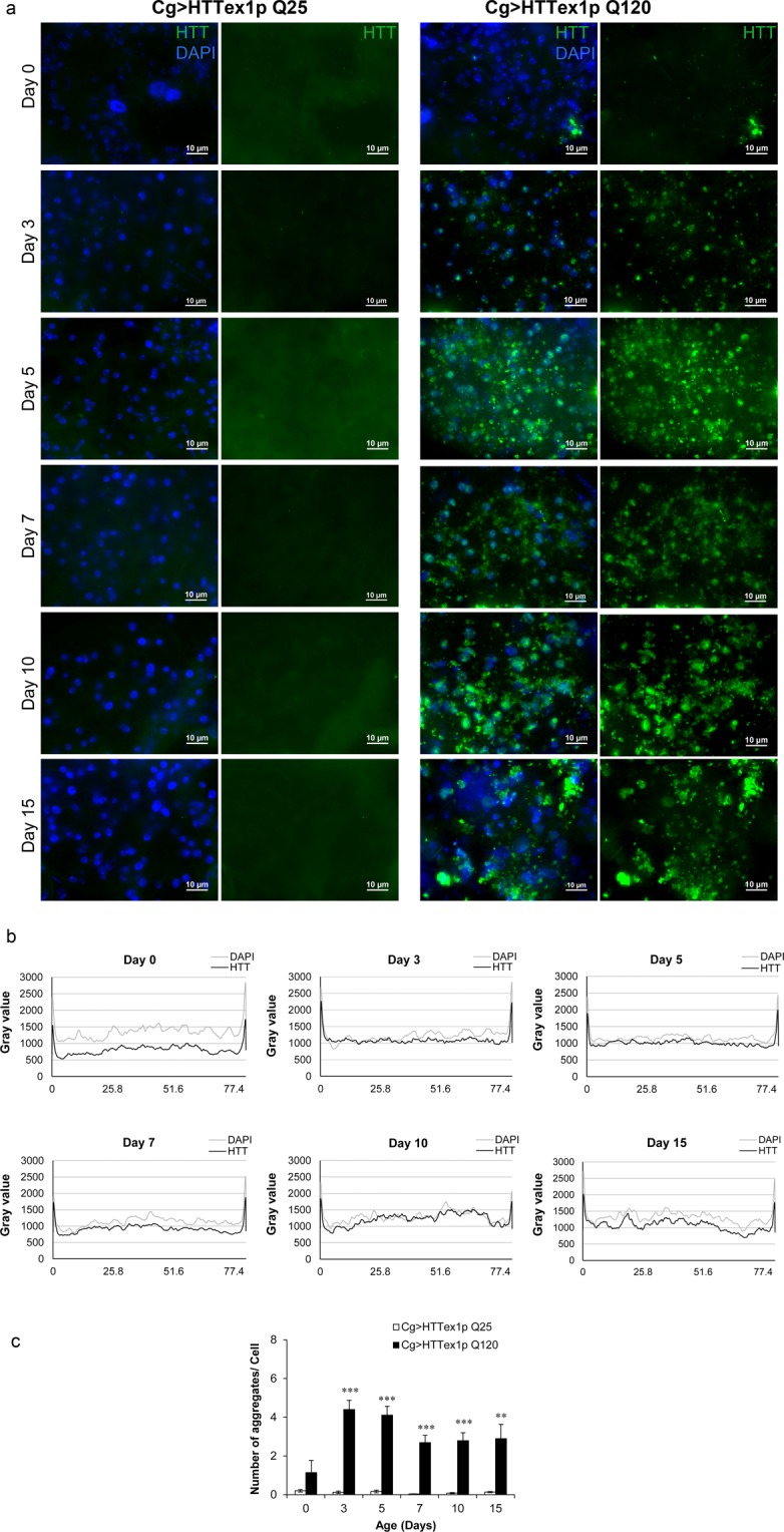
Aggregation of mHTT in the abdominal FB in vivo. Abdominal FB dissected from 25QHTTex1 and 120QHTTex1 females at different ages were probed with anti-HTT antibody to reveal the accumulation and/or aggregation of HTT. (a) Cg-GAL4 driven-mHTT exon1 peptides form aggregates in the abdominal FB. At day3, aggregates begin to appear as bright puncta (green) in 120QHTTex1 flies (right panel). Along with the aggregates, soluble diffused forms are also detected in the FB at this time point. On the other hand, FB of 25QHTTex1 does not show any accumulation of unexpanded HTT exon1 throughout the indicated ages (left panel). (n = 6/genotype/age) Scale bar, 10 µm. (b) The image analysis of the FB from 120QHTTex1 flies (a, right panel) is shown as intensity profiles of HTT (black) and DAPI (gray) at different ages. The intensity of HTT signal is lowest at day0 and higher at day10 while the intensity of DAPI signal is relatively constant throughout. (c) The number of detectable aggregates per cell was determined by analysing the 3D stacks of all the images in (a) using the ImageJ software (n = 6/genotype/age). The objects i.e. apparent aggregates are detected strongly in the FB from 120QHTTex1 flies (a, right panel) while the FB from 25QHTTex1 generates negligible signal (a, left panel). The number of objects detected in the day0 old FB from 120QHTTex1 flies specimens is notably less than the number of objects in the following days.
