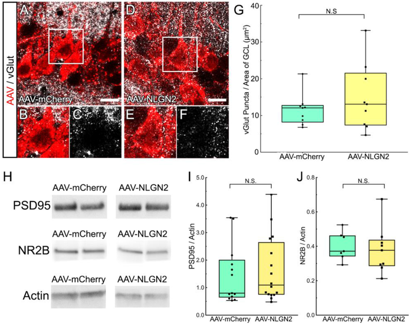
Figure 5. Excitatory synaptic proteins vGlut, PSD-95, and NR2B were not altered after transduction of hippocampal neurons with AAV-NLGN2.
A, Representative confocal photomicrographs showing putative sites of synaptic contacts indicated by vGlut+ puncta (white) on AAV-mCherry+ pyramidal neurons (red). B, C, Magnified views of selected cells. D, Confocal images of vGlut+ puncta (white) apposed to AAV-NLGN2+ pyramidal neurons (red). E, F, Magnified views of selected cells. G, Quantification for vGlut+ puncta/μm2 of mCherry immunofluorescence showing that vGlut+ puncta were not increased in neurons transduced with the AAV-NLGN2 vector, compared to AAV-mCherry vector (p=0.5631, Student’s t-test). H, Representative Western blots comparing PSD95 and NR2B protein bands in hippocampal membrane fractions after transduction with the AAV-NLGN2 or the AAV-mCherry vectors. I, Quantification of protein bands revealed that PSD95 was not significantly different in the tissue transduced with the two different vectors (p=0.6622, Student’s t-test). J, Membrane levels of NR2B were also comparable in hippocampal tissue transduced with the AAV-NLGN2 or AAV-mCherry vectors (p=0.7113, Student’s t-test). A, D, Scale bars equal 20 microns.

