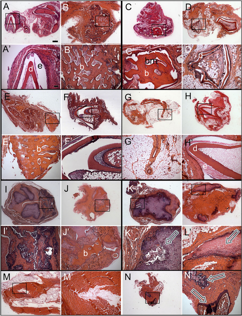
Figure 5.

Histological analyses of subcutaneous post-natal recombinant constructs. Haematoxylin and eosin (H&E) stained paraffin embedded and sectioned implants. Letter panels with primes represent higher magnification images of the boxed regions of each corresponding lettered panel. (A,A’) Cusp formation in a pDE-T + pDM-T sample harvested after 1 month. (B,B’) Bone/osteodentin-like tissue in a pDE-T + pDM-T sample after 3 months. (C,C’) Bone/osteodentin-like tissue in a pDE-T + hDM sample after 1 month. (D,D’) Cusp formation in a pDE-T + hDM sample after 3 months. (E,E’) Bone/osteodentin-like tissue in a pDE-T + S sample after 1 month. (F,F’) Cusp formation in a pDE-T + S sample after 3 months. No calcified tissue was found in pDE-C + pDM-C samples at 1 month (G), and only in one implant at 3 months (H). Bone/osteodentin-like tissue present in hGE-C + pDE-T samples at 1 month (I) and 3 months (J). (K,L) Necrotic DM tissue was observed in most pDM-T + S implants at 1 month (K) and 3 months (L) (arrows). (M,N) Most hGE-C + hDM-C implants showed no sign of calcification (M), but contained mucosal epithelial-like tissues (N). hDM-C, human dental pulp derived dental mesenchymal cells; pDE-T, porcine dental epithelial tissue; pDM-T, porcine dental mesenchymal tissue; S, acellular collagen scaffold. Scale bars: A–N, 500 μm; A’-N’, 50 μm.
