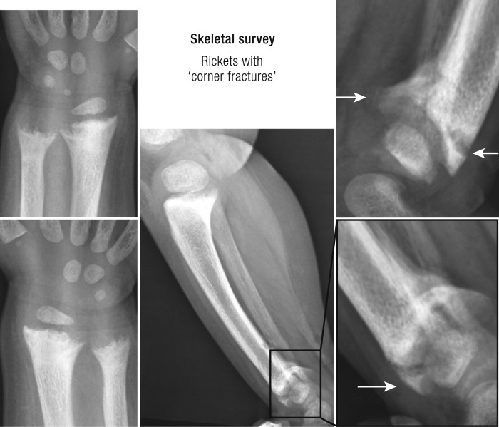
Figure 2.
Radiologic image of nutritional rickets. A radiologic image of a 19-mo-old child with nutritional rickets is shown. The child was born from Indian parents, living in Australia, after a normal pregnancy of 40 wk and received exclusive breastfeeding for 18 mo without vitamin D supplementation. Height and weight were around the 50th percentile. Medical attention was asked because of genu varum and delayed walking. Serum calcium (2.01 mmol/L; 2.10 to 2.65) and phosphate were slightly decreased. Serum 25OHD was <18 nmol/L and alkaline phosphatase and PTH (126 pmol/L; 1.0 to 7.0) levels were high.

