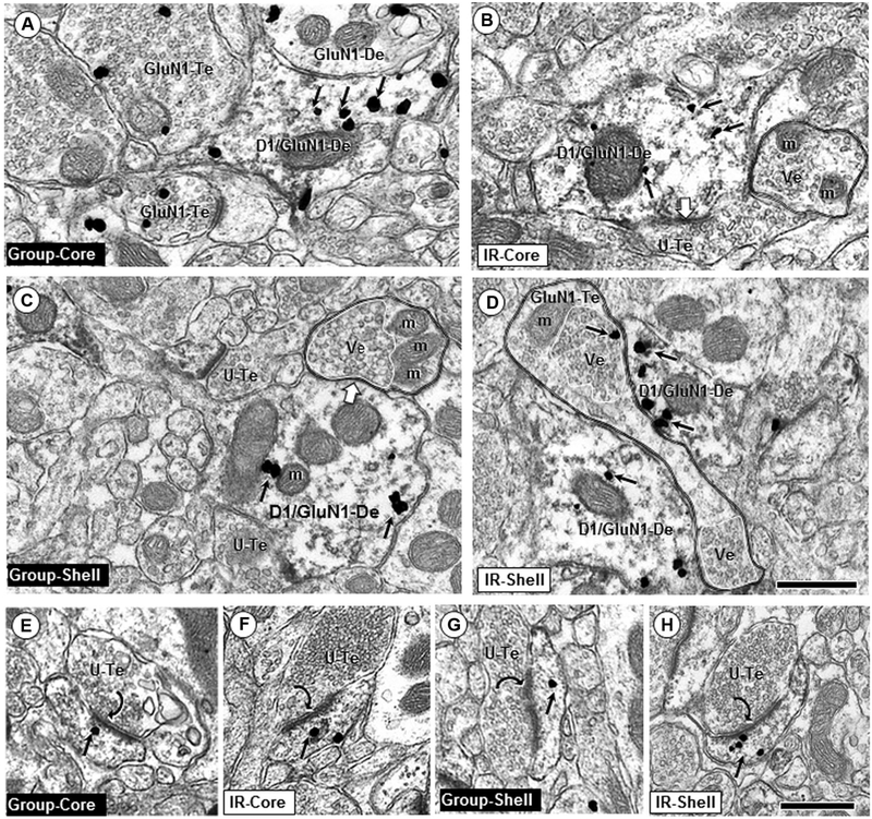
Fig. 3.
Dendritic shaft and spine profiles dual labeled for GluN1 and D1. In dendritic shafts, GluN1 immunogold is frequently located near endomembranes (em) and mitochondria (m) and less commonly on non-synaptic portions of the plasma membranes in profiles from group-reared (a, c) and IR (b, d). Axon terminals apposing these dendrites are often unlabeled (U-Te; b, c) but sometimes also contain GluN1 immunogold (GluN1-Te; a, d). Unlabeled axon terminals identified by double black lines show partially segregated compartmental distributions of densely packed vesicles (Ve, white line) and mitochondria (m). In D, two dually labeled dendrites are contacted by an “en passant” axon terminal (GluN1-Te) with two clusters of vesicles (see Gracy and Pickel 1996 for comparison with dopamine axons in the Acb shell). Note the qualitative decrease of GluN1 immuno-gold (small arrows) in IR rats in dendritic shafts co-expressing D1Rs (D1/GluN1-De) in the Acb core (a, b), but not in dually labeled dendritic shafts in the shell (c, d). e–h show dually labeled spines in core or shell of IR and control adult rats; scale bar = 500 nm

