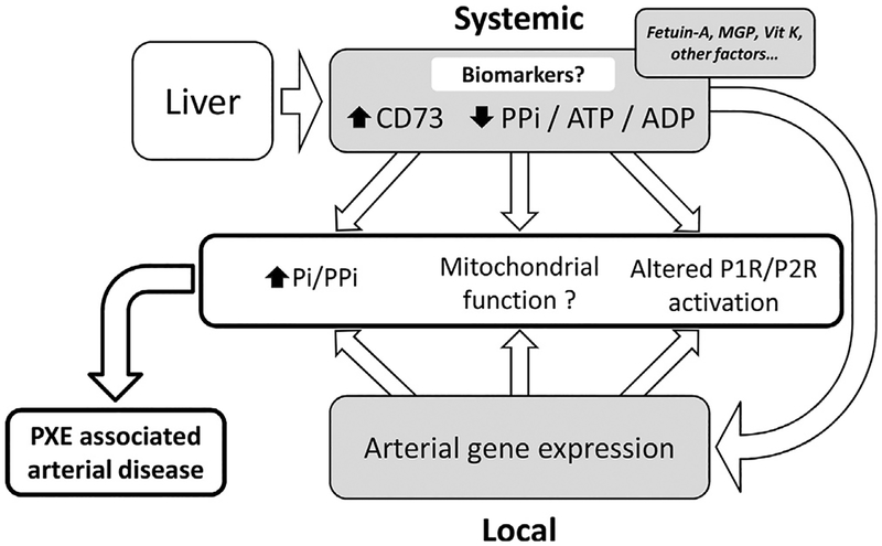
Figure 5. Proposal of a summary diagram for the pathophysiology of PXE.
In the context of liver (central) ABCC6 deficiency, plasma levels of PPi and adenine nucleotides (ATP, ADP) are decreased and 5′-nucleotidase activity is increased each of these, representing potential disease biomarkers. ABCC6 deficiency is also associated with modifications of purine and phosphate gene expression in (remote) affected tissues like arteries. Both systemic and local alterations in purine and phosphate metabolism contribute to PXE-associated peripheral arterial disease. ADP, adenosine diphosphate; ATP, adenosine triphosphate; MGP, matrix gla protein; Pi, inorganic phosphate; PPi, pyrophosphate; PXE, pseudoxanthoma elasticum; Vit, vitamin.

