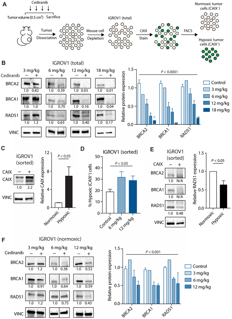
Fig. 1. Cediranib down-regulates HDR factor expression in ovarian tumor xenografts in both hypoxia-dependent and hypoxia-independent pathways.
(A) Schematic of mouse treatment, isolation of human IGROV1 cancer cells from mouse stroma, and FACS-based separation of hypoxic and normoxic cells from IGROV1 tumors. (B) Western blot of HDR factors in IGROV1 ovarian cancer xenograft tumors in mice treated with vehicle or the indicated doses of cediranib [two-way analysis of variance (ANOVA), effect of cediranib: P < 0.0001; control, n = 14; 3 mg/kg, n = 2 (pooled to one sample); 6 mg/kg, n = 9; 12 mg/kg, n = 3; 18 mg/kg, n = 2 tumors]. (C) Western blot of CAIX in sorted normoxic (CAIX−) and hypoxic (CAIX+) cells isolated from IGROV1 tumors (unpaired t test, P < 0.05; n = 5 tumors). (D) Percentage of hypoxic tumor cells after cediranib treatment (one-way ANOVA, P < 0.05; control, n = 14; 6 mg/kg, n = 10; 12 mg/kg, n = 3 tumors). (E) Western blot of HDR factors in sorted normoxic and hypoxic cells isolated from IGROV1 tumors (unpaired t test, P < 0.05; n = 3 tumors). (F) Western blot of HDR factors in sorted normoxic cells isolated from IGROV1 tumors in control [dimethyl sulfoxide (DMSO)] and cediranib-treated mice at the indicated doses [two-way ANOVA, effect of cediranib: P < 0.001; control, n = 9; 3 mg/kg, n = 2 (pooled to one sample); 6 mg/kg, n = 5; 12 mg/kg, n = 3 tumors]. Data are represented as means ± SEM. Numbers below Western blot panels represent relative quantification of the respective bands normalized to loading control by densitometry.

