
Figure 5.
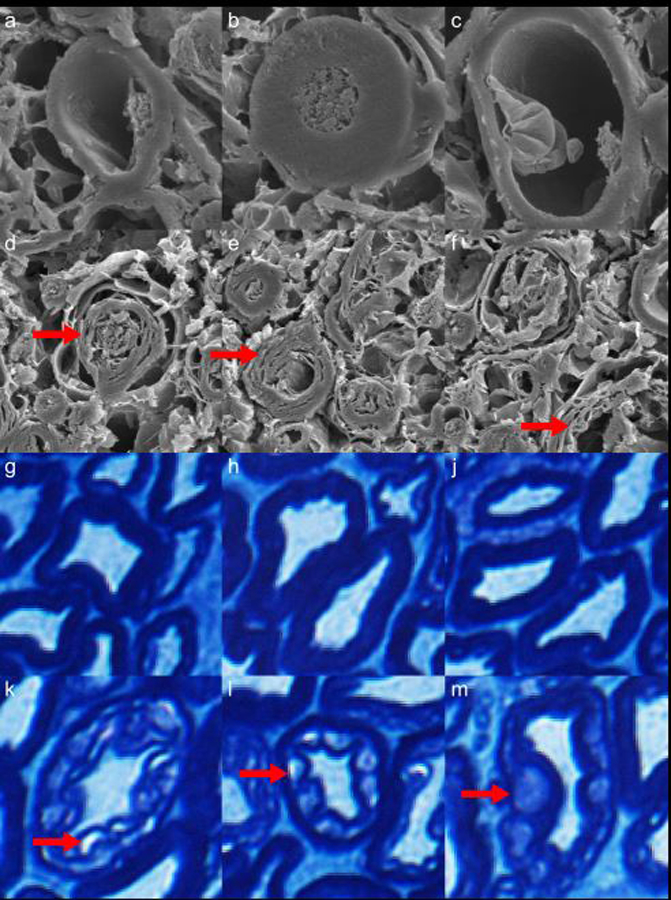
Electron microscopy (a-f) and Luxol fast blue-stained optical microscopy (g-m) images of myelinated axons from a rhesus macaque cervical spinal cord specimen fixed in formalin for 6 months. In the second (d-f) and fourth (k-m) rows, vacuoles caused by inadequate fixation by immersion in formalin are indicated by red arrows. These vacuoles may be linked to the observed increases in F and MWF in fixed tissue, relative to tightly-wrapped myelin sheathes (a-c, g-j). Note: Luxol fast blue stains phospholipids, which are present in all cell membranes.