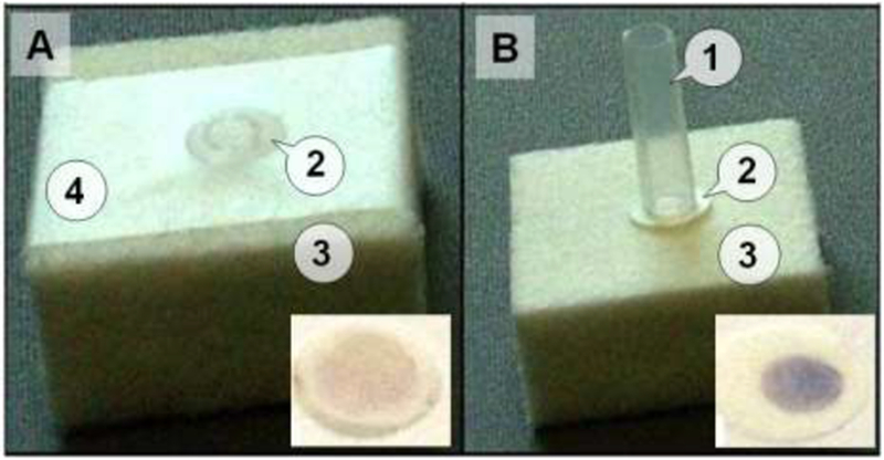
Figure 2: Laboratory setups of the FTCA cassette.

Panel A: A conventional system with retainer filter (membrane) attached with tape (4) on the absorbent pad. The sample is applied directly on part of the membrane through an opening in the tape. Panel B: The FTCA cassette prototype comprised of a sample delivery tube/cylinder (1) held over the membrane either manuall y or by mechanical means; WBC retainer filter (membrane, 2); and an absorbent pad (3). The insets show images of the membranes with final results obtained from a whole blood sample after the color development step.
