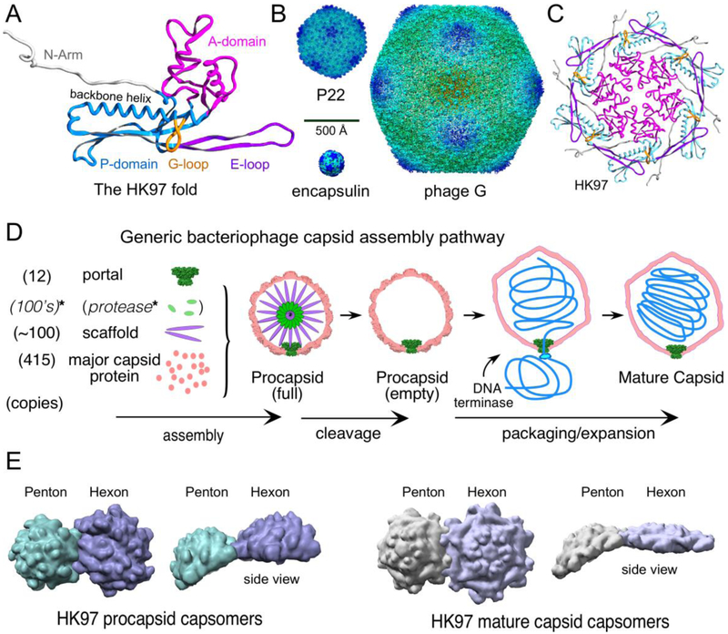
Figure 1. Components and features of capsids.
A. The HK97 fold with important common features labeled and color-coded as indicated in the figure. The ribbon diagram is from the mature HK97 capsid chain A (PDB 1OHG). B. The size range of the HK97 fold. Shown are images of surface rendered cryo-EM density maps from the T=1 encapsulin of Thermotoga maritima [6], the P22 mature capsid [19], and Bacillus megaterium jumbophage G [7]. C. A hexon from the HK97 mature capsid colored as in (A). D. A generic pathway for capsid assembly that applies, in general features, to nearly all double-stranded DNA tailed phages and Herpesviruses. *Note that while a capsid maturation protease is a common feature, there are many bacteriophages that do not utilize one. E. Examples of the shape changes of capsomers that occur when procapsids convert to mature procapsids. Surface rendered images are shown of the hexons and pentons of phage HK97 from X-ray structures (PDB ID 3E8K (procapsid) and 1OGH (mature)). Structural models in figures were visualized using Chimera [47] or SPDBV [48].

