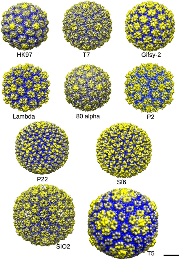
Figure 3. Procapsid gallery.
Ten examples of procapsid structures from the Protein Data Base (PDB) or Electron microscopy database (EMDB) rendered at low resolution using Chimera [47] so that the shapes of the procapsid hexons can be discerned. Procapsids are round (not angular) and have lumpy capsomers with a domed and asymmetric shape, as noted in the text. The procapsids are radially cued with yellow being the furthest from and blue being closest to the center of each particle. The scale bar represents 20 nm. Shown are the procapsids of phages HK97 (PDB 3E8K, [17]); T7 (EMD-1321 ,[51]); Gifsy-2 (EMD-1691, [52]); Lambda (EMD-1507, [53]); 80alpha (EMD-7030, PDB 6B0X;[42]); P2 (EMD-5406, [54]); P22 (EMD-5149, [15]); Sf6 (EMD-5724, [55]); SIO2 (EMD-5383, [56]); and T5 (not deposited, [57]).

