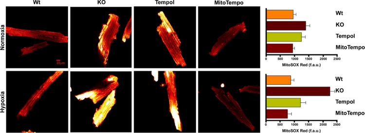
Figure 6. MitoTempo and Tempol decreases elevated O2.− levels in gKO myocytes after hypoxia/reoxygenation.
Global Trpm2 KO myocytes were pre-incubated with MitoTempo (50 nM) or Tempol (100 μM) or vehicle for 60 min before subjected to 30 min of normoxia (21% O2-5% CO2) or hypoxia (1% O2-5% CO2) followed by 30 min of reoxygenation. During reoxygenation, myocytes were loaded with the mitochondrial O2.− sensitive fluorophore MitoSOX Red (Materials and Methods). Representative confocal images are shown for WT, gKO, gKO-Tempol and gKO-MitoTempo after normoxic (upper panel) and H/R (bottom panel) incubations. There are 9 WT, 15 gKO, 15 gKO-Tempol and 15 gKO-MitoTempo myocytes incubated under normoxic conditions and 12 WT, 15 gKO, 12 gKO-Tempol and 15 gKO-MitoTempo myocytes incubated under hypoxic conditions followed by reoxygenation. Under normoxic conditions, there are no statistically significant differences in O2.− levels among WT, gKO, gKO-Tempol and gKO-MitoTempo myocytes although gKO myocytes tended to have higher O2.− levels compared to WT myocytes (Upper panel). After H/R, KO myocytes had significantly (p<0.001) higher superoxide levels than WT myocytes. Both MitoTempo and Tempol were effective in reducing the elevated O2.− levels in gKO-H/R myocyte (p<0.001 compared to gKO-H/R myocytes).

