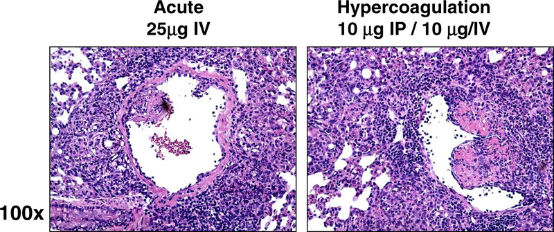
Figure 3. Inflammation Accompanied with Vascular Occlusion in the Hypercoagulation Model Post TDM Administration.
Acute IV administration reveals accumulation of monocytes and lymphocytes surrounding vascular beds, but limited inflammation in subendothelial regions. In contrast, the hypercoagulation model led to aggressive inflammation throughout the parenchmya, with alterations to subendothelial structure lining vasculature. Formalin fixed lung sections; H&E stain; representative sections, 100x magnification. Representative sections from repeated experiments; 4–6 mice per group per experiment.

