
Fig. 5.
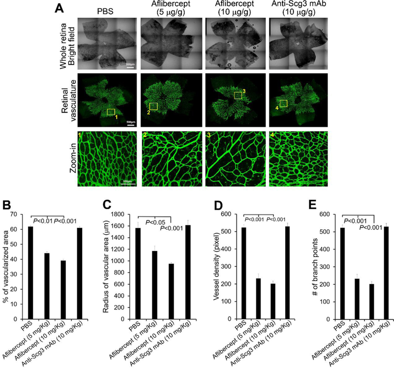
Aflibercept, but not anti-Scg3 mAb, suppresses the development of retinal vasculature at P6. Aflibercept (5 or 10 mg/Kg body weight) or anti-Scg3 ML78.2 mAb (10 mg/Kg) were injected i.p. into mice at P1, 3 and 5. At P6, retinas were isolated, stained with fluorescent isolectin B4, analyzed by confocal microscopy. a Representative images of retinas in the bright and fluorescent fields. Bar= 500 μm (top two rows), 100 μm (bottom row). b Percentage of vascularized area. c Radius of vascularized retina. d Branch points. e Vessel density. n=5 eyes (PBS), 6 eyes (aflibercept 5 or 10 mg/Kg), n= 4 eyes (anti-Scg3 mAb). ± SEM, one-way ANOVA test.