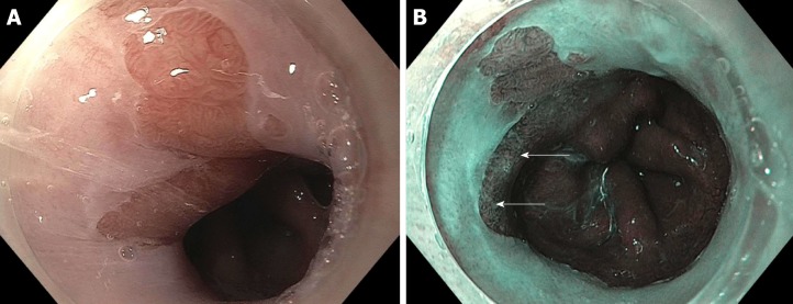
Figure 1.
A patient with Barrett’s and high-grade dyspalsia with narrow band imaging. A: A segment of Barrett’s esophagus on high definition white light endoscopy (HDWLE); B: narrow band imaging (NBI) from a patient with prior long segment disease post two sessions of endoscopic resection, 4 sessions of radiofrequency ablation, and one session of cryotherapy. The HDWLE did not show any features concerning for dysplasia. The NBI shows an area of disrupted vessels (upper white arrow, lower white arrow) concerning for dysplasia.

