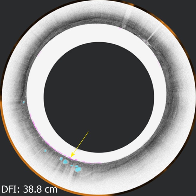
Figure 3.

Volumetric laser endomicroscopy from the same patient showing cross-sectional view of the area of overlap (yellow arrow 5.73” 5.41”) between three features of dysplasia (orange is lack of layering, blue is glandular structures, and pink is a hyper-reflective surface).
