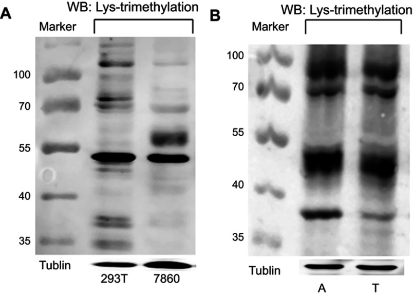
Figure 1.
Lys-trimethylation was observed in different proteins in kidney-derived cells and tissues.
Notes: (A) The levels of Lys-trimethylation in HEK293T and 786-0 cells were quantified by Western blotting; (B) The levels of Lys-trimethylation in kidney cancer tissues and adjacent normal renal cortex tissues were quantified by Western blotting. Tubulin served as the loading control. Marker: protein marker (Fermentas, USA, Catalog # 26616-ladder-002); The targeted proteins were stained with fluorescence-conjugated secondary antibodies (IRDye800CW or IRDye680 conjugated IgG, LICOR).
Abbreviations: 293T, HEK293T cells; 7860, 786-0 cells; A, adjacent normal renal cortex tissues; T, kidney cancer tissues.

