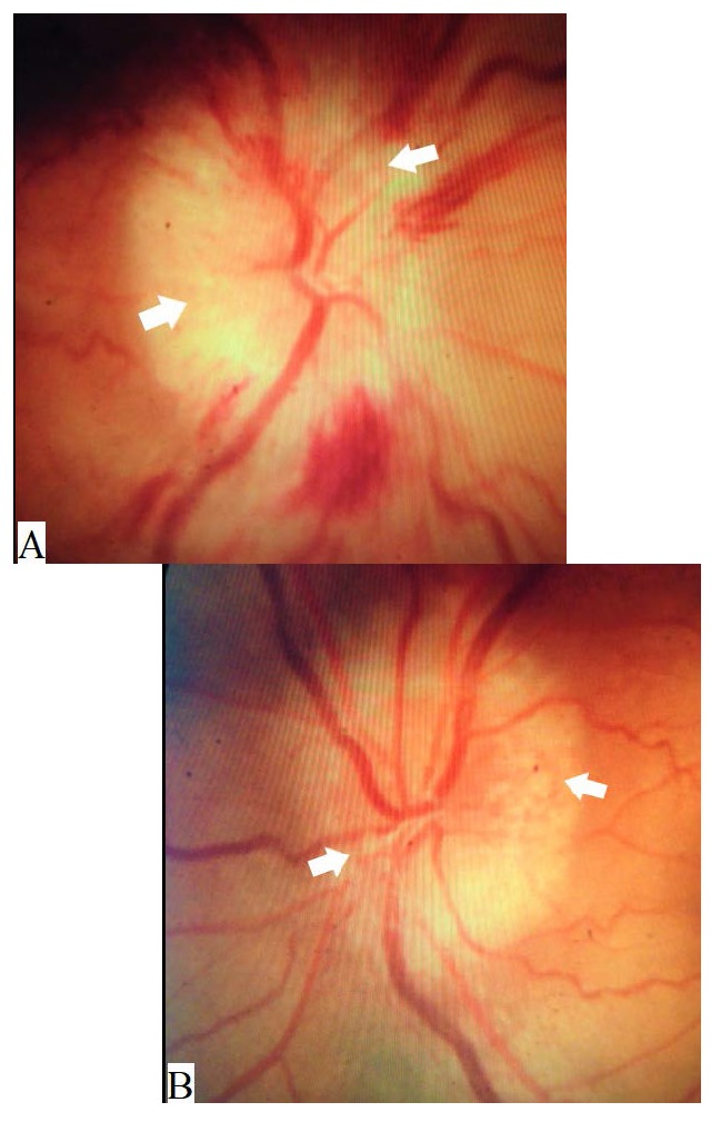
Fig. 1.

Color photography of the optic discs showing swelling of both optic discs with blurred margins more temporal than nasal and multiple peculiar nodules (white arrows) suggestive of optic disc drusen, with multiple flame shaped hemorrhages over the right disc (A) and an anomalous left retinal vasculature (B)esence of a large “cotton-ball” colony in the patient’s right eye
