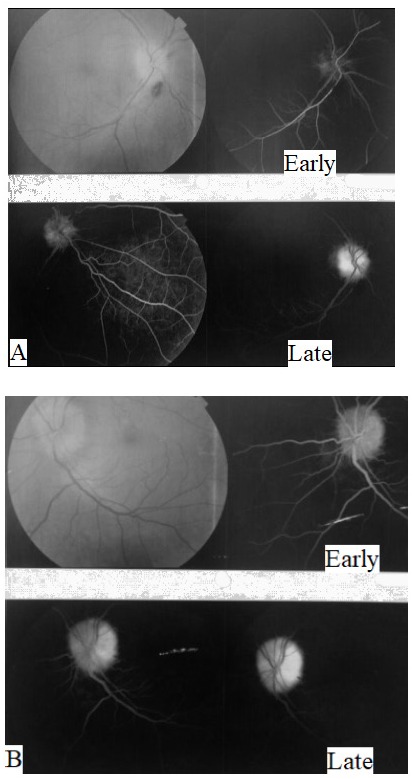
Fig. 3.

Fundus photography and fluorescein angiography of both eyes. A. The right optic disc was swollen with flame shaped hemorrhages, and fluorescein angiography showed early disc hypofluorescence indicating hypoperfusion with late staining and mild leakage. B. The left optic disc was swollen with anomalous retinal vasculature, and fluorescein angiography showed early disc hyperfluorescence with late staining and no leakage side
