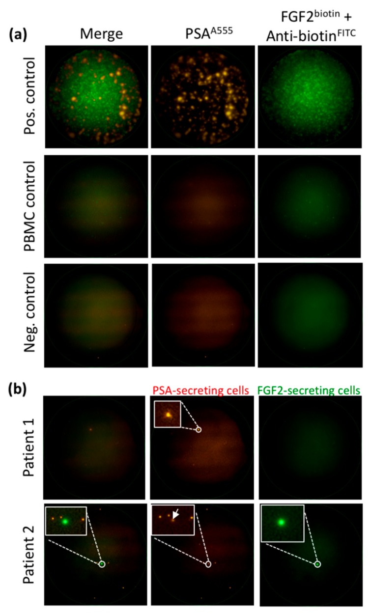
Figure 3.
Detection of viable CTCs using the dual fluorescent EPISPOTPSA/FGF2 assay. (a) Positive and negative controls. LNCaP (shown in figure) and NBTII cells that secrete PSA and FGF2, respectively, were used as positive controls (2000 cells/well), whereas wells with peripheral blood mononuclear cells (PBMC) and without cells were used as negative controls. Each immuno-spot corresponds to the protein “fingerprint” of one viable cell. (b) Patient samples. PSA-secreting cells are considered as CTCs in blood samples from patients with PCa (Patient 1). A subset of CTCs can secrete FGF-2 in addition to PSA (Patient 2). Representative images of PSA-positive, FGF2-positive and double PSA/FGF2 immuno-spots (merge) corresponding to viable CTCs. Immuno-spots were detected and observed using the C.T.L. Elispot Reader, 50× magnification.

