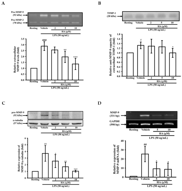
Figure 1.
Effect of HA on MMP-9-mediated gelatinolysis and expression induced by LPS. THP-1 cells (5 × 10⁵ cells/0.5 mL) were dispensed onto 24-well plates and treated with LPS (50 ng/mL) for 24 h. Cells were treated with the indicated concentrations of HA (2, 5 and 10 μM) or vehicle for 15 min before treatment with a stimulant. Cell-free supernatants were then assayed for MMPs and TIMP-1 activity by gelatin zymography (A) and reverse zymography (B). THP-1 cells (106 cells/mL) were dispensed onto 6-well plates and were treated with LPS (50 ng/mL) for 24 h (C) or 8 h (D) at the indicated concentrations of HA or vehicle for 15 min before treatment with LPS. Cell lysates were obtained and analyzed for MMP-9 protein expression by Western blotting or for MMP-9 mRNA expression by RT-PCR. Data represent means ± S.D. from three independent experiments. # p < 0.05, ## p < 0.01 and ### p < 0.001 as compared with the resting, * p < 0.05, ** p < 0.01 and *** p < 0.001 as compared with the vehicle.

