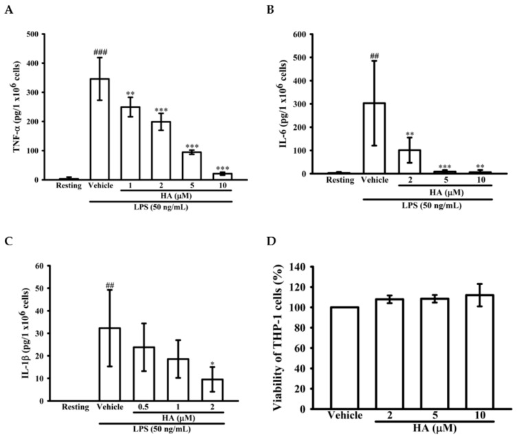
Figure 2.
Effect of HA on pro-inflammatory cytokines production induced by LPS in THP-1 cells without cellular toxicity. THP-1 cells (5 × 10⁵ cells/0.5 mL) were dispensed onto 24-well plates and were treated with the indicated concentrations of HA or vehicle for 15 min followed by treatment with LPS (50 ng/mL) for 4 h (A) and 24 h (B,C). Cell-free supernatants were then assayed for the level of TNF-α (A), IL-6 (B) and IL-1β (C) by ELISA. (D) THP-1 cells treated with the indicated concentrations of HA or vehicle for 24 h. Cell viability was quantified by the ability of mitochondria to reduce the tetrazolium dye MTT in viable cells. Data represent means ± S.D. from three independent experiments. ## p < 0.01 and ### p < 0.001 as compared with the resting; * p < 0.05, ** p < 0.01 and *** p < 0.001 as compared with the vehicle.

