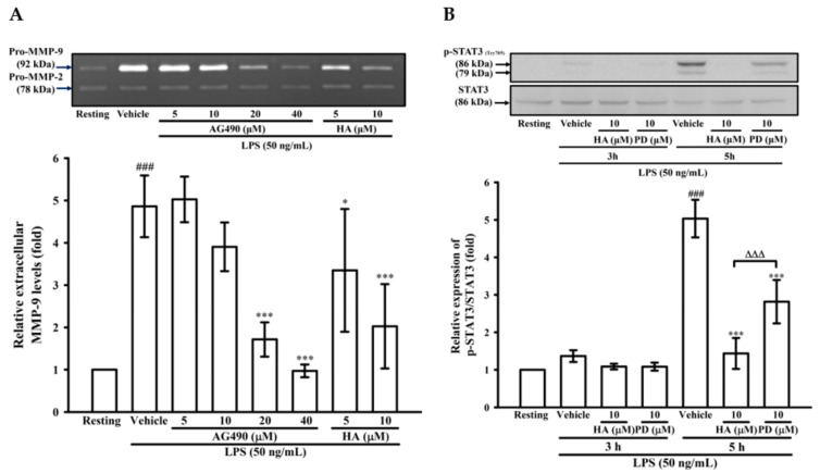
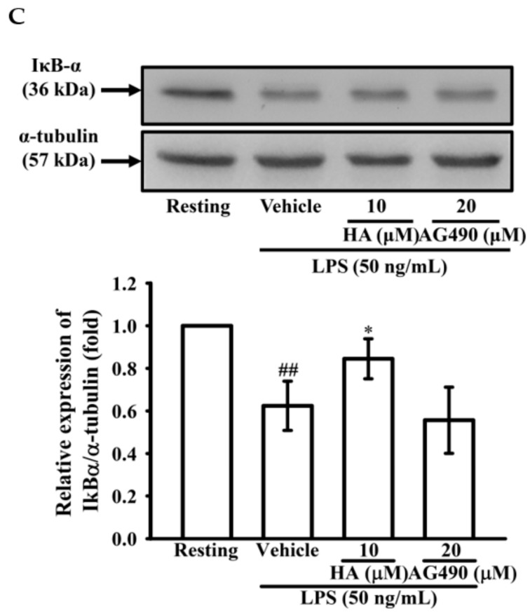
Figure 4.
Effect of JAK2-STAT3 cascade inhibition on LPS-mediated MMP-9 expression. THP-1 cells (5 × 10⁵ cells/0.5 mL) were dispensed onto 24-well plates and treated with LPS (50 ng/mL) for 24 h. Cells were treated with the indicated concentrations of AG490 (5, 10, 20, and 40 μM), HA (5 and 10 μM) or vehicle for 15 min before treatment with LPS. Cell-free supernatants were then assayed for MMPs activity by gelatin zymography (A). THP-1 cells (106 cells/mL) were dispensed onto 6-well plates and were treated with LPS (50 ng/mL) for 3 h, 5h (B) and 60 min (C) at the indicated concentrations of HA, PD (10 μM), AG490 (10 μM) or vehicle for 15 min before treatment with LPS. Cell lysates were obtained and analyzed for STAT3 (Tyr705) phosphorylation and IκBα degradation by Western blotting. Data represent means ± S.D. from three to four independent experiments, respectively. ## p < 0.01 and ### p < 0.001 as compared with the resting; * p < 0.05 and *** p < 0.001 as compared with the vehicle; ∆∆∆ p < 0.001 as compared with the group pretreated with HA pre-treatment.


