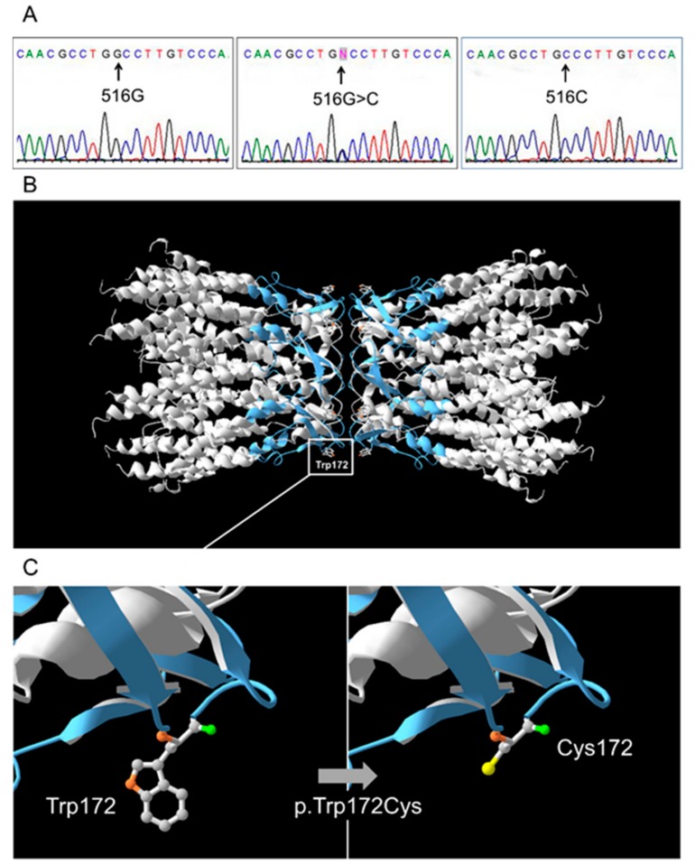
Figure 1.
(A) Identification of the c.516G>C (p.Trp172Cys) mutation by Sanger sequencing. (B) The 3D structure of normal protein connexin 26 (Cx26-WT). Two adjacent Cx26-WT hemichannel subunits with designation of Trp (tryptophan) on position 172. The extracellular loop of Cx26 (E2, 155-192 amino acid residues) where variant p.Trp172Cys located is marked by blue. (C) Close-up view of Cx26-WT (Trp172) and mutant Cx26-p.Trp172Cys (Cys172) connexin 26.

