Figure 3.
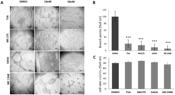
Effect of HDACis on U87MG cells ability to form vascular tubes. (A) Representative images of vascular tubes formed by U87MG cells in control group (DMSO 0.1%) and in treated groups with HDACis at final concentrations of 10 and 50 nM. Network of tubes, in each well, were analyzed directly under an inverted microscope with 10× phase contrast and imaged. Scale bars: 50 µm. (B) Quantification of vascular branching in control group and groups treated with 50 nM HDACis. Branch point (sites of intersection of at least three tubes) number from five random fields in each well was counted and expressed as a percentage of branch points formed in the control sample, taken as 100%. As shown, TSA, MS275, SAHA and MC1568 HDACis strongly inhibit U87MG cells ability to form vascular tubes at 50 nM. (C) The cell rate viability assayed by MTT assay. At concentration of 50 nM, no significant differences are detected in treated groups as compared to the control group in 24 h. *** p-value < 0.001.

