Figure 6.
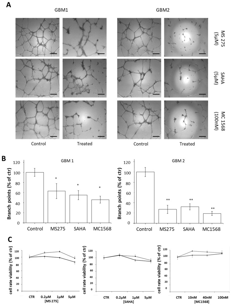
Effect of HDACis on the ability of CSCs isolated from two human GBMs to form vascular tubes. (A) Representative images of vascular tubes formed by GBM1 and GBM2 CSCs treated with vehicle (DMSO 0.1%) or with the tested drugs, at the indicated concentrations. Network of tubes, in each well, were directly analyzed under an inverted microscope with 5× phase contrast and imaged. Scale bar: 250 μm. (B) Quantification of vascular branching of A. Branch point number in each well was counted and expressed as a percentage of branch points formed in the control sample, taken as 100%. As shown, 24 h of treatment with MC1568, SAHA and MS275 reduce CSCs ability to form vascular tubes, with higher efficacy in GBM2 cells. (C) The cell viability rate, assayed by MTT reduction assay, evaluated in GBM CSCs; no significant differences are detected in treated groups as compared to the control group after 24 h of treatment. * p-value < 0.05, ** p-value < 0.01.

