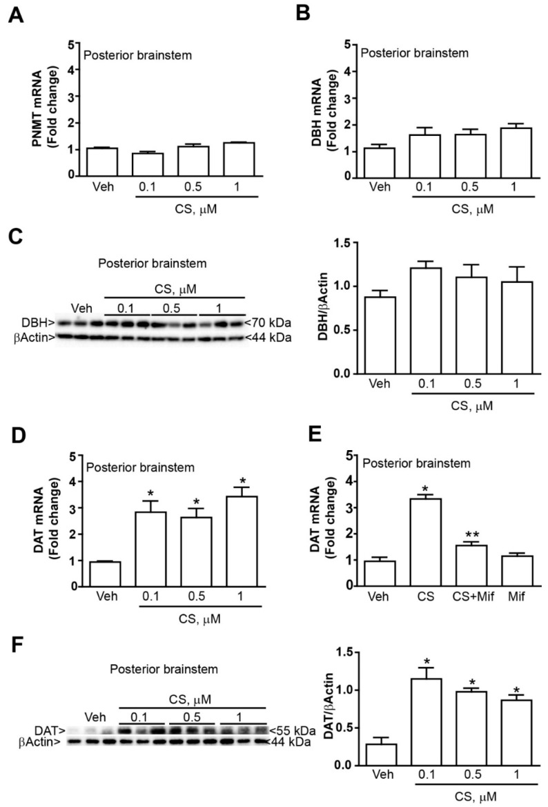
Figure 4.
Corticosterone increases DAT expression without modifying DBH and PNMT expression in the mouse brainstem. Real time PCR of (A) the adrenergic marker PNMT and (B) the noradrenergic marker DBH within the caudal part of the brainstem at 24 h incubation with either vehicle (Veh, ethanol 0.01%) or corticosterone (CS, 0.1, 0.5 or 1 µM). (C) Western blot of DBH within the caudal part of the brainstem at 24 h incubation with either vehicle (Veh, ethanol 0.01%) or corticosterone (CS, 0.1, 0.5 or 1 µM). (D) Real time PCR of the dopaminergic marker (DAT) within the caudal part of the brainstem at 24 h incubation with either vehicle (Veh, ethanol 0.01%) or corticosterone (CS, 0.1, 0.5 or 1 µM). (E) Real time PCR of the DAT within the caudal part of the brainstem at 24 h incubation with either vehicle or CS (0.1 µM), in the presence or absence of the selective glucocorticoid receptors antagonist mifepristone (Mif, 10 µM). (F) Western blot of DAT within the caudal part of the brainstem at 24 h incubation with either vehicle (Veh, ethanol 0.01%) or corticosterone (CS, 0.1, 0.5 or 1 µM). The lines of the βActin are the same showed in the Figure 2E as the western blot for DAT was performed by incubating with the anti-DAT primary antibody the very same membrane used for TH immunoblotting. All values are expressed as the means±SEM. * p < 0.05 compared with the vehicle. ** p < 0.05 compared with the vehicle and CS.

