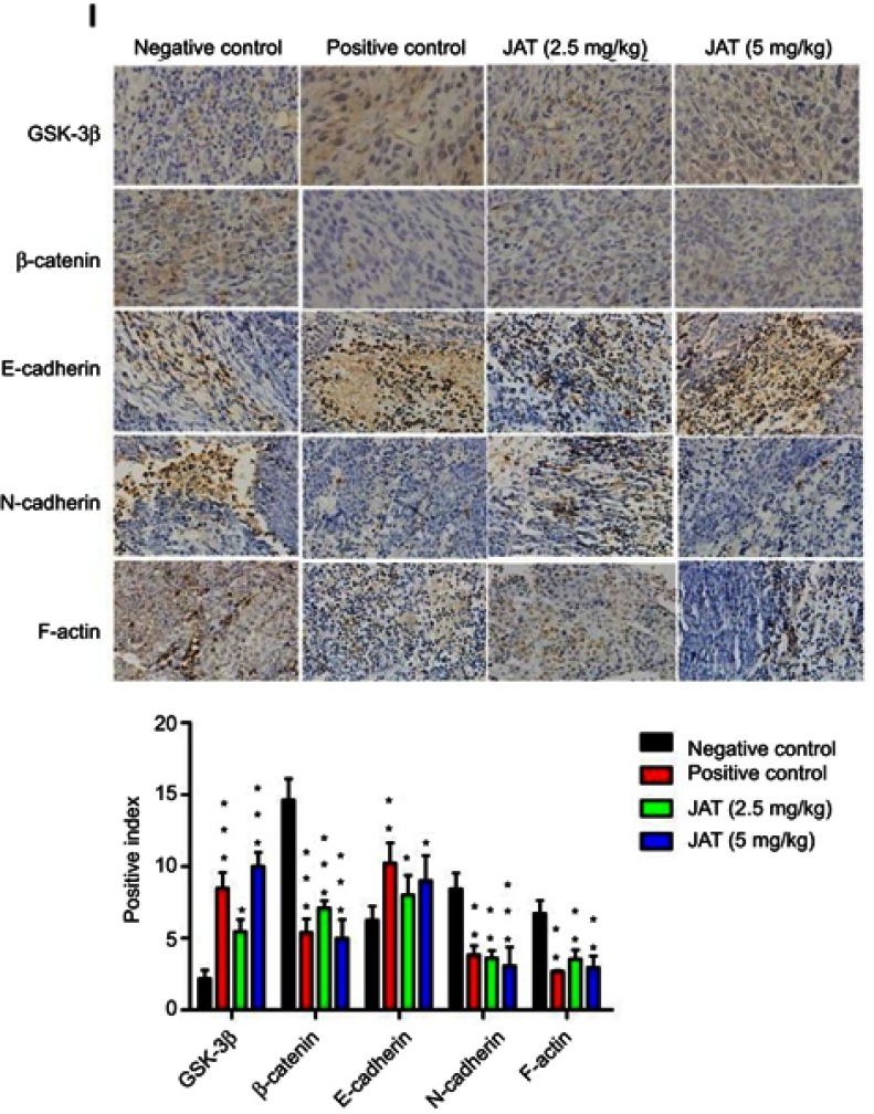
Figure 8.
JAT suppressed tumor growth and metastasis in vivo.
Notes: (A) Tumor volume was measured every 5 days for 30 days. (B) Tumor weight was measured after the experiment. (C) The survival rate of mice was shown by the Kaplan–Meier survival curves. (D) Mice body weight was measured every 5 days for 30 days. (E) Mice were sacrificed at 35 d, removed the liver and kidney tissues and weighed, calculated their organ index. (F and G) Histological analysis of different tumor and lung tissues was detected by H&E staining (×400). (H) The apoptosis of different tumor tissues was monitored by TUNEL method (×400). The bright areas represent apoptotic cells. (I) The positive expression of GSK-3β, β-catenin, E-cadherin, N-cadherin, and F-actin in tumor tissues was detected by immunohistochemistry (×400). Immunohistochemistry results were analyzed by Image J software. All values were presented as the mean±standard deviation (SD). *P<0.05, **P<0.01, and ***P<0.001 compared with negative control group.
Abbreviation: JAT, jatrorrhizine.

