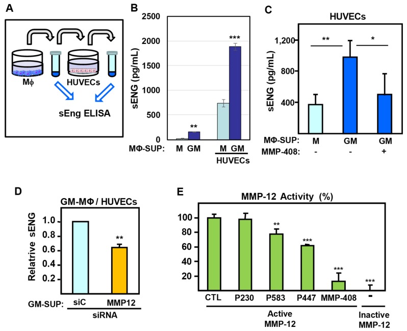
Figure 6.
Secretion of endothelial soluble endoglin induced by MΦ supernatants. (A–D) As indicated in the scheme (A), culture supernatants (SUP) from GM-MΦ (GM) or M-MΦ (M) were added to monolayers of HUVECs. After 24 h incubation, the culture media were collected, and the sEng content was measured by ELISA in all samples. The values of sEng were calculated after subtracting the basal release of sEng present in HUVECs supernatants (B–D). (B) The levels of sEng from GM-MΦ and M-MΦ supernatants (left two bars) are compared to those after incubation with HUVECs (right two bars). (C) Soluble endoglin levels from GM-MΦ and M-MΦ supernatants after incubation with HUVECs in the presence or absence of MMP-408. (D) Soluble endoglin levels from supernatants of GM-MΦ, previously transfected with siRNA-MMP-12 or -control (siC), after incubation with HUVECs. (E) Effect of endoglin peptides (P230, P583, P447) and the specific inhibitor MMP-408 on the proteolytic activity of recombinant human MMP-12. Control samples with vehicle (CTL), the GL-less peptide P230, as well as an inactive MMP-12, are included. The MMP-12 activity was measured using the fluorogenic substrate Mca-PLGL-Dpa-ARNH2, and the statistical significance respect to the control sample (CTL) is indicated. Statistical analysis (n = 3) was calculated using the ANOVA test, and data are presented as mean ± SD. (*p < 0.05; **p < 0.01; ***p < 0.001).

