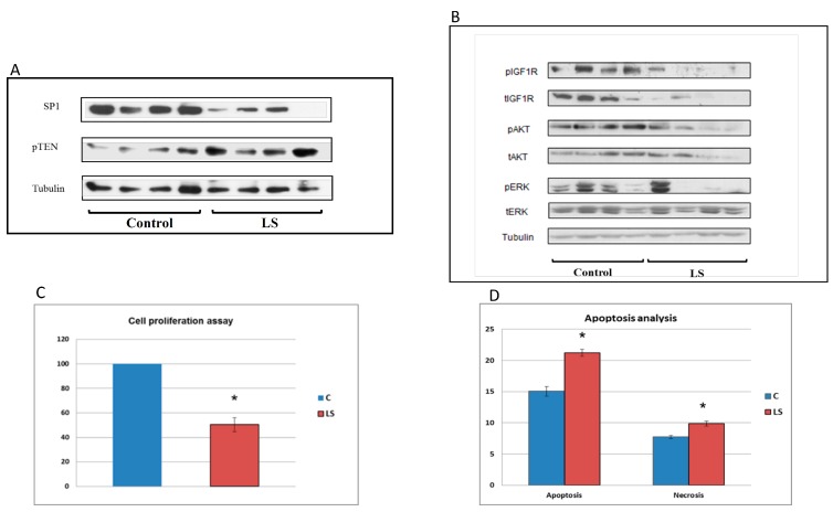
Figure 4.
Analysis of signaling pathways associated with cancer protection in LS. (A) Western blot analysis of Sp1 and pTEN levels in LS-derived and control lymphoblastoids. Lymphoblastoid cell lines of four LS patients and four controls were lysed and extracts were electrophoresed through SDS-PAGE. Blots were incubated with antibodies against Sp1 and pTEN. The lanes correspond to individual controls and patients. (B) Western blot analysis of downstream mediators of IGF1 action in LS. Cell extracts were resolved on SDS-PAGE and membranes were incubated with antibodies against phospho- and total-IGF1 receptor (IGF1R), phospho- and total-AKT and phospho- and total-ERK. Tubulin levels were measured as a loading control. (C) Cell proliferation of LS and control cells. Proliferation of LS- and control-derived lymphoblastoid cells was assessed using an XTT colorimetric kit. The statistical significance of differences between groups was assessed by Student’s t-test. Legend: *, significantly different versus control (p < 0.05); red bars, LS; blue bars, controls. (D) Basal apoptosis and necrosis of LS and control cells. Apoptosis and necrosis were measured by flow cytometry analysis after staining cells with an annexin-V antibody and propidium iodide (PI). Necrotic cells were stained with PI as well as annexin V; apoptotic cells were stained only with annexin V. The figure was adapted from [55].

