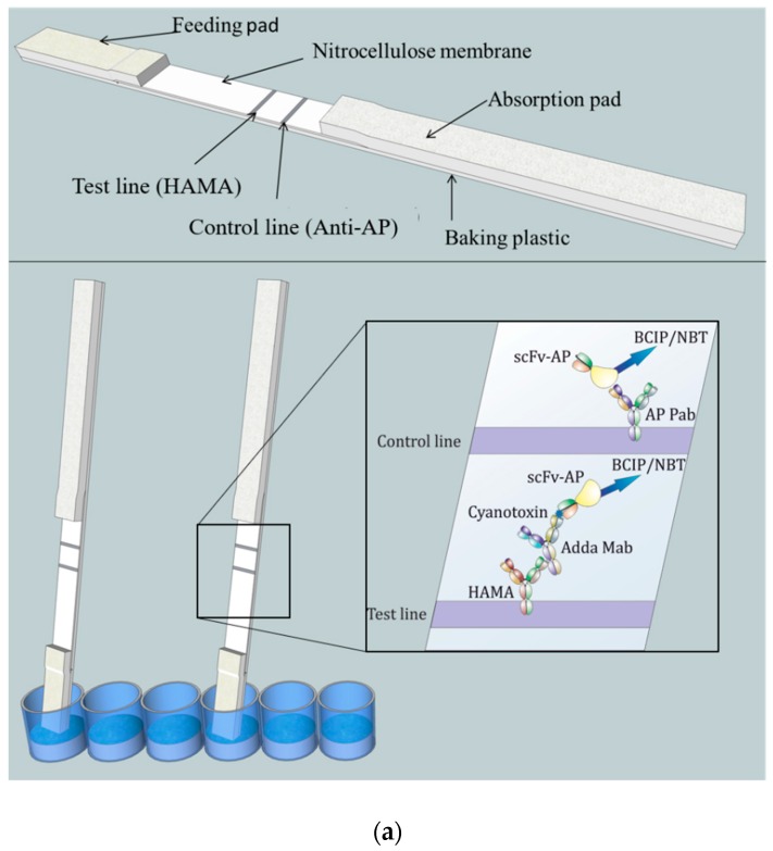
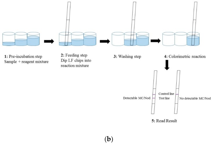
Figure 1.
(a) Conceptual illustration of the noncompetitive chromogenic LFIA. Top: The structure of the LFIA strip comprising feeding pad, nitrocellulose membrane, and absorbtion pad. Test line (HAMA: human anti-mouse antibody) and control line (anti-AP Pab) were printed on nitrocellulose membrane. Bottom: Principle of the chromogenic anti-IC LFIA. The sample absorbing procedure was performed on microtiter well. Once the liquid is absorbed, the reagents move along the immunostrip by capillary action and respective antibody components attach to the test and control lines. In the presence of MC/Nod in the sample the anti-Adda Mab:MC/Nod:anti-IC-scFv-AP complex become captured by HAMA on the test line through the Fc portion of the anti-Adda Mab. The excess unbound anti-IC-scFv-AP continue to migrate until captured by the anti-AP Pab in the control line. The alkaline phosphatase (AP) fused with the anti-IC scFv triggers an enzymatic reaction producing a chromogenic precipitant in the presence of BCIP/NBT substrate. In the absence of MC/Nod, anti-IC scFv-AP does not bind to the test line and continue to migrate until captured in the control line. The control line ensures that the chromogenic reaction is functional in the LFIA. (b) The main steps of the LFIA procedure where the sample absorption took place on microtiter well. The assay protocol comprised of four main steps. (1) In the preincubation step, sample and reagents are mixed together to allow formation of the anti-Adda Mab:MC/Nod:anti-IC-scFv-AP complex in the presence of toxin. (2) In the feeding step, LF chips were dipped into the pre-incubated reaction mixture through feeding pad where antibody components or any formed immunocomplex migrated through test and control line. (3) The washing step removes any unbound antibody components. (4) In the fourth step, colorimetric reaction took place in the presence of chromogenic substrate. (5) Finally, the developed color on the strips can be observed by naked eye or through instrument (optional). MC = microcystin; Nod = nodularin.


