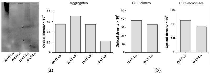
Figure 3.
(a) Membrane image of CML western blot measured of BLG heated in the presence of lactose (La) in a wet system (W) or a dry system (D) at low temperatures (LT) or high temperatures (HT). G90: soy protein extract glycated with glucose for 90 min (positive control). Numbers indicate aggregates (1), BLG dimers (2), and BLG monomers (3). (b) Optical density of bands visible on the CML western blot categorized in high-MW aggregates, BLG dimers, and BLG monomers as indicated on the membrane.

