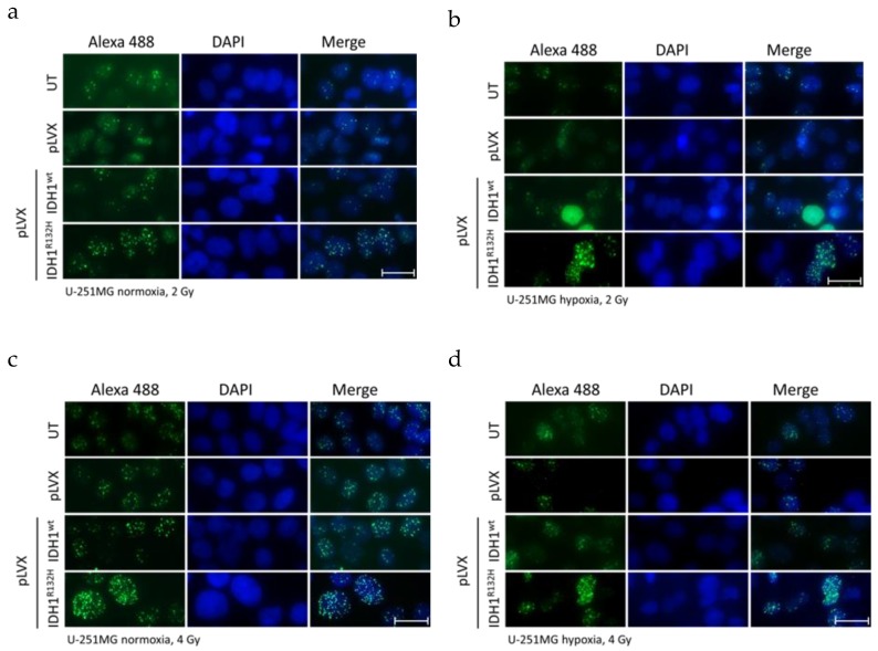
Figure 1.
Effect of isocitrate dehydrogenase 1 (IDH1)R132H gene expression on the accumulation of residual γH2AX foci after radiation in U-251MG cells. Representative immunofluorescence images of γH2AX of U-251MG cells 24 h after irradiation with 2 (a,b) and 4 Gy (c,d) under normoxia (a,c) and hypoxia (b,d). Green: γH2AX foci; blue: Cell nuclei (DAPI). n = 3 independent experiments; scale bar = 25 µm. Normoxia (21% O2), hypoxia (< 0.1% O2). UT: untreated, pLVX: cells stably transduced with empty vector, pLVX IDH1wt: IDH1wt-expressing cells, pLVX IDH1R132H: IDH1R132H-expressing cells.

