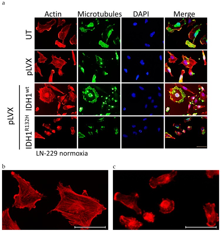
Figure A5.
Immunofluorescence staining of transduced LN-229 glioma cells stably expressed IDH1wt or IDH1R312H protein. (a) Representative immunofluorescence staining of actin stress fibers and microtubules in LN-229 cells using phalloidin-TRITC and anti-tubulin antibody. Immunofluorescence staining was achieved 24 h after seeding. Cell nuclei were counterstained with DAPI. n = 3 independent experiments; scale bar = 50 µm. (b) Enlarged representative immunofluorescence staining as a representative example for untreated cells, empty vector cells (pLVX) and IDH1wt cells; the enlarged part is taken from pLVX cells; scale bar = 50 µm. (c) Enlarged representative immunofluorescence staining as a representative example for untreated cells, empty vector cells (pLVX) and IDH1R132H cells; the enlarged part is taken from IDH1R132H cells; scale bar = 50 µm. UT: untreated, pLVX: cells stably transduced with empty vector, pLVX IDH1wt: IDH1wt-expressing cells, pLVX IDH1R132H: IDH1R132H-expressing cells.

