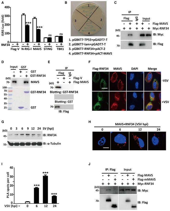
Luciferase activity driven by the ISRE promoter in HEK293T cells transfected with Myc‐RNF34 and Flag‐V, Flag‐N‐RIG‐I, Flag‐MAVS, Flag‐STING, or Flag‐TBK1. Luciferase assays were performed 24 h after transfection.
Y2H analysis in the AH109 yeast strain co‐transformed with the indicated plasmids. A positive RNF34‐MAVS interaction resulted in colony formation on synthetic medium lacking tryptophan, leucine, adenine, and histidine containing X‐gal. pGBKT7‐TP53 + pGADT7‐T and pGBKT7‐lam+pGADT7‐T were used as positive and negative controls, respectively. AH109 co‐transfected with pGBKT7‐RNF34 + pACT‐2 was used to exclude the self‐activation of RNF34.
Immunoprecipitation analysis of HEK293T cells transfected with Myc‐RNF34 and Flag‐MAVS or Flag‐V. Anti‐Flag or IgG agarose immunoprecipitates were analyzed using immunoblotting with an anti‐Myc or anti‐Flag antibody.
GST‐tagged RNF34 was subjected to a pull‐down assay with HEK293T cell lysates. Immunoblot with an anti‐MAVS antibody is shown in the top panel. Loading of the GST proteins assessed using Coomassie blue staining is shown in the bottom panel. GST was used as a negative control.
Anti‐Flag or IgG immunoprecipitates prepared from cells transfected with Flag‐MAVS or Flag‐vector‐expressing plasmids were subjected to SDS–PAGE and blotted onto a nitrocellulose membrane. The nitrocellulose membrane was incubated with soluble GST‐RNF34 (upper panel) or GST (middle panel) for 2 h and then analyzed with anti‐Flag antibody.
Representative confocal images of immunofluorescence staining for Flag‐RNF34 colocalization with endogenous MAVS in THP‐1 cells infected with VSV for 12 h. Scale bar, 10 μm.
Immunoblot showing the levels of the RNF34 protein in THP‐1 cells infected with VSV (MOI = 1.0) for the indicated times. α‐Tubulin was used as a loading control.
In situ PLA assay of the RNF34‐MAVS complex in HEK293T cells infected with VSV (MOI = 1.0) for the indicated times using an anti‐RNF34 or anti‐MAVS antibody. RNF34‐MAVS complex, red; nuclei, blue. Scale bar, 5 μm.
One hundred cells in Fig
2H were counted, and the quantification of PLA signals per cell is shown.
Immunoprecipitation analysis of HEK293T cells transfected with Myc‐RNF34 and Flag‐MAVS or Flag‐mMAVS. Anti‐Flag immunoprecipitates were analyzed using immunoblotting with anti‐Myc or anti‐Flag antibody.
Data information: Cell‐based studies were performed independently at least three times with comparable results. The luciferase reporter and ELISA data are presented as means ± SEM. Two‐tailed Student's
< 0.001.

