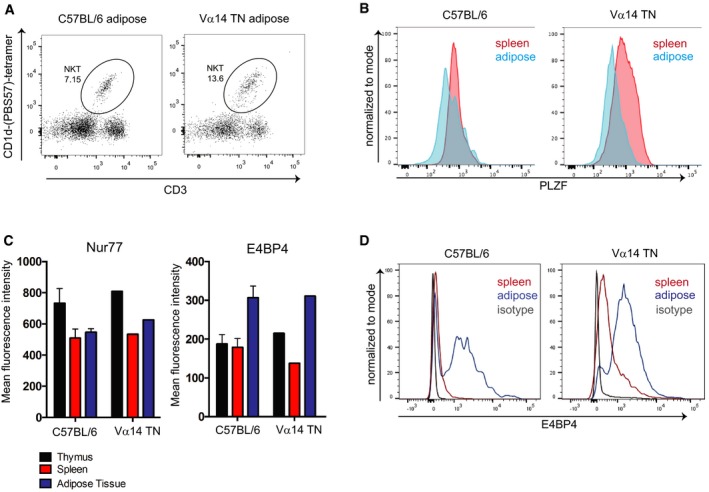
Figure EV2. Adipose iNKT cells from Vα14 TN mice are indistinguishable from C57BL/6‐derived adipose iNKT cells.

- Flow cytometry analysis of iNKT cell abundance in white adipose tissue from a Vα14 TN mouse.
- Spleen cells and stromal vascular fractions of white adipose tissue from Vα14 TN or C57BL/6 mice were stained intracellularly with anti‐PLZF and analyzed by flow cytometry. Histograms shown are gated on CD1d‐(PBS57)‐tetramer+ CD3+ cells.
- Thymus, spleen, and adipose tissue were harvested from C57BL/6 mice and Vα14 TN mice. Cell suspensions were stained with antibodies to CD3, Nur77, E4BP4, and CD1d‐(PBS57)‐tetramer and analyzed by flow cytometry. Mean fluorescence intensity of Nur77 and E4BP4 staining after gating on iNKT cells is shown. N = 3 per group. Error bars are SEM.
- Representative histograms of E4BP4 staining are shown. Plots are gated on total CD3+ cells.
