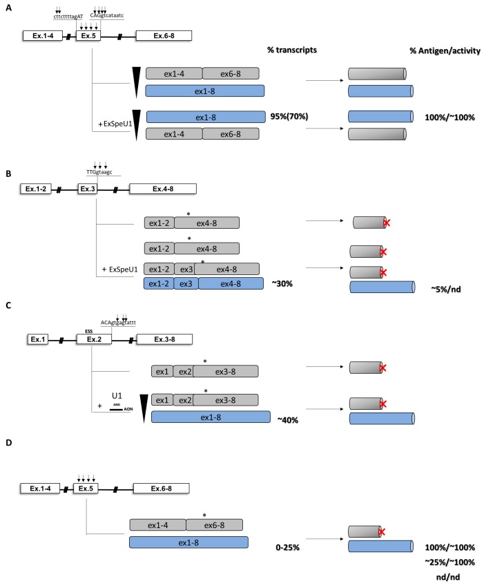
Figure 6.
U1-mediated rescue in hemophilia B. Hemophilia B models caused by multiple nucleotide changes at the 3’ss (exon 5, A), at the 5’ss (exons 2, 3 and 5, B, C and D) or within exon (exon 5, panel D) of various exons of the F9 gene. Schematic representation of the genomic context (left panel), splicing transcripts (middle panel) and protein isoforms (right panel) is reported. Sequences of the splice sites and position of mutations (black arrows) are indicated. Frameshift of the coding sequence and premature stop codons are reported by asterisks and red X letter, respectively. Percentages of transcripts, antigen and relative coagulation activity are reported. Values in rounded brackets indicate experiments in mouse model of the disease.

